

What is a choledochal cyst? A choledochal cyst is a rare congenital condition where cysts form in the bile ducts. These ducts carry bile from the liver to the small intestine, aiding digestion. When cysts develop, they can cause blockages, leading to serious complications like jaundice, pancreatitis, or even liver damage. Symptoms often include abdominal pain, nausea, and yellowing of the skin. Early diagnosis is crucial for effective treatment, which usually involves surgical removal of the cysts. Understanding this condition can help in recognizing symptoms early and seeking timely medical intervention.
Key Takeaways:
- Choledochal cysts are rare, but they can cause serious complications if not treated. Early detection and surgical removal are crucial for a good prognosis.
- Symptoms of choledochal cysts include jaundice, abdominal pain, and an enlarged liver. Minimally invasive surgery and regular follow-ups can lead to positive long-term outcomes.
What is a Choledochal Cyst?
A choledochal cyst is a rare congenital condition involving cystic growths in the bile ducts. These cysts can cause various complications if not treated. Here are some intriguing facts about choledochal cysts.
- Choledochal cysts are more common in females than males, with a ratio of 3:1.
- They are most frequently diagnosed in East Asian populations.
- The condition is usually present at birth but may not be detected until later in life.
- Symptoms often include jaundice, abdominal pain, and an enlarged liver.
- There are five types of choledochal cysts, classified based on their location and structure.
- Type I cysts are the most common, accounting for about 80-90% of cases.
- Type II cysts are diverticulum-like outpouchings of the bile duct.
- Type III cysts, also known as choledochoceles, are located within the duodenal wall.
- Type IV cysts involve multiple cysts in both intrahepatic and extrahepatic bile ducts.
- Type V cysts, also known as Caroli disease, are characterized by cystic dilations of the intrahepatic bile ducts.
Causes and Risk Factors
Understanding the causes and risk factors of choledochal cysts can help in early detection and management. Here are some key points.
- The exact cause of choledochal cysts is unknown.
- Genetic factors may play a role in the development of these cysts.
- An abnormal pancreaticobiliary junction is often associated with choledochal cysts.
- This abnormal junction allows pancreatic enzymes to reflux into the bile ducts, causing inflammation and cyst formation.
- Family history of choledochal cysts can increase the risk.
- Environmental factors have not been conclusively linked to the development of choledochal cysts.
- Some studies suggest a possible link between choledochal cysts and certain infections during pregnancy.
- Biliary atresia, a condition where bile ducts are blocked or absent, can sometimes be associated with choledochal cysts.
- Choledochal cysts are more common in children than adults.
- Early diagnosis and treatment are crucial to prevent complications.
Symptoms and Diagnosis
Recognizing the symptoms and understanding the diagnostic process is essential for timely intervention. Here are some important facts.
- Jaundice, a yellowing of the skin and eyes, is a common symptom.
- Abdominal pain, especially in the upper right quadrant, is frequently reported.
- Nausea and vomiting can occur due to bile duct obstruction.
- Fever may indicate an infection in the bile ducts.
- An enlarged liver, or hepatomegaly, can be a sign of choledochal cysts.
- Ultrasound is often the first imaging test used to detect choledochal cysts.
- Magnetic Resonance Cholangiopancreatography (MRCP) provides detailed images of the bile ducts.
- Endoscopic Retrograde Cholangiopancreatography (ERCP) can be used for both diagnosis and treatment.
- Blood tests may show elevated liver enzymes and bilirubin levels.
- A liver biopsy is rarely needed but can help in complex cases.
Treatment and Management
Effective treatment and management are vital to prevent complications. Here are some key aspects.
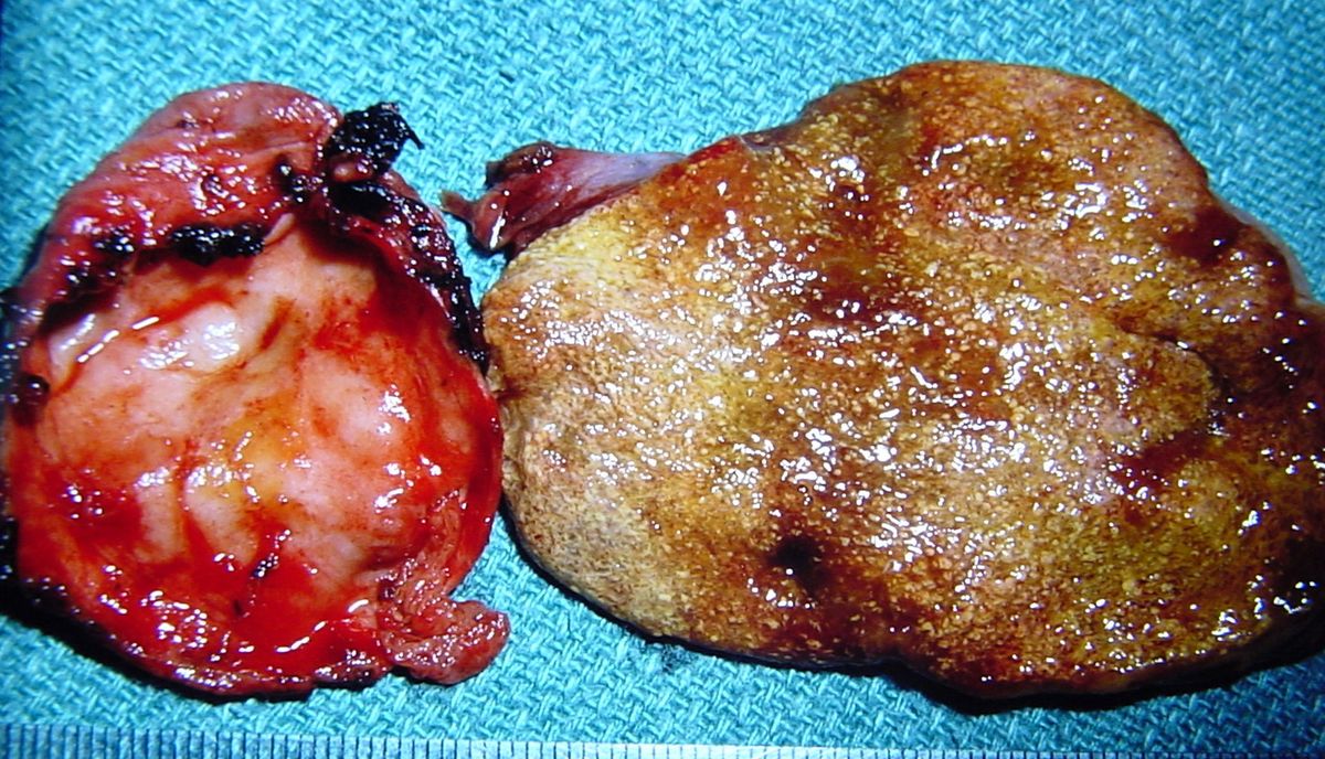
- Surgical removal of the cyst is the primary treatment.
- Complete excision of the cyst reduces the risk of cancer.
- Hepaticojejunostomy is a common surgical procedure used to reconstruct the bile ducts.
- Laparoscopic surgery offers a minimally invasive option for cyst removal.
- Post-surgery, patients may need regular follow-ups to monitor liver function.
- Antibiotics may be prescribed to prevent or treat infections.
- Pain management is an important aspect of post-surgical care.
- In some cases, liver transplantation may be necessary.
- Early intervention can significantly improve the prognosis.
- Long-term outcomes are generally good with appropriate treatment.
Complications and Prognosis
Understanding potential complications and the overall prognosis helps in managing expectations and planning treatment. Here are some critical points.
- Untreated choledochal cysts can lead to cholangitis, an infection of the bile ducts.
- Pancreatitis, inflammation of the pancreas, is another possible complication.
- Biliary cirrhosis, a severe liver condition, can develop if bile flow is obstructed.
- Choledochal cysts increase the risk of bile duct cancer, also known as cholangiocarcinoma.
- Gallstones may form within the cysts or bile ducts.
- Liver abscesses, or pockets of infection, can occur.
- Portal hypertension, increased blood pressure in the liver's portal vein, is a potential risk.
- With timely and appropriate treatment, most patients have a good prognosis and can lead normal lives.
Final Thoughts on Choledochal Cysts
Choledochal cysts, though rare, can have significant impacts on health. Understanding their symptoms, causes, and treatment options is crucial for early detection and management. These cysts often present in childhood but can also appear in adults. Common symptoms include abdominal pain, jaundice, and fever. Early diagnosis through imaging techniques like ultrasound and MRI can lead to effective treatment, often involving surgical removal.
Ignoring symptoms can lead to complications like cholangitis, pancreatitis, or even bile duct cancer. Awareness and timely medical intervention are key. If you or someone you know experiences persistent abdominal pain or jaundice, consult a healthcare professional.
Stay informed, stay proactive, and prioritize your health. Knowledge about choledochal cysts can make a difference in outcomes, ensuring better health and peace of mind.
Frequently Asked Questions
Was this page helpful?
Our commitment to delivering trustworthy and engaging content is at the heart of what we do. Each fact on our site is contributed by real users like you, bringing a wealth of diverse insights and information. To ensure the highest standards of accuracy and reliability, our dedicated editors meticulously review each submission. This process guarantees that the facts we share are not only fascinating but also credible. Trust in our commitment to quality and authenticity as you explore and learn with us.