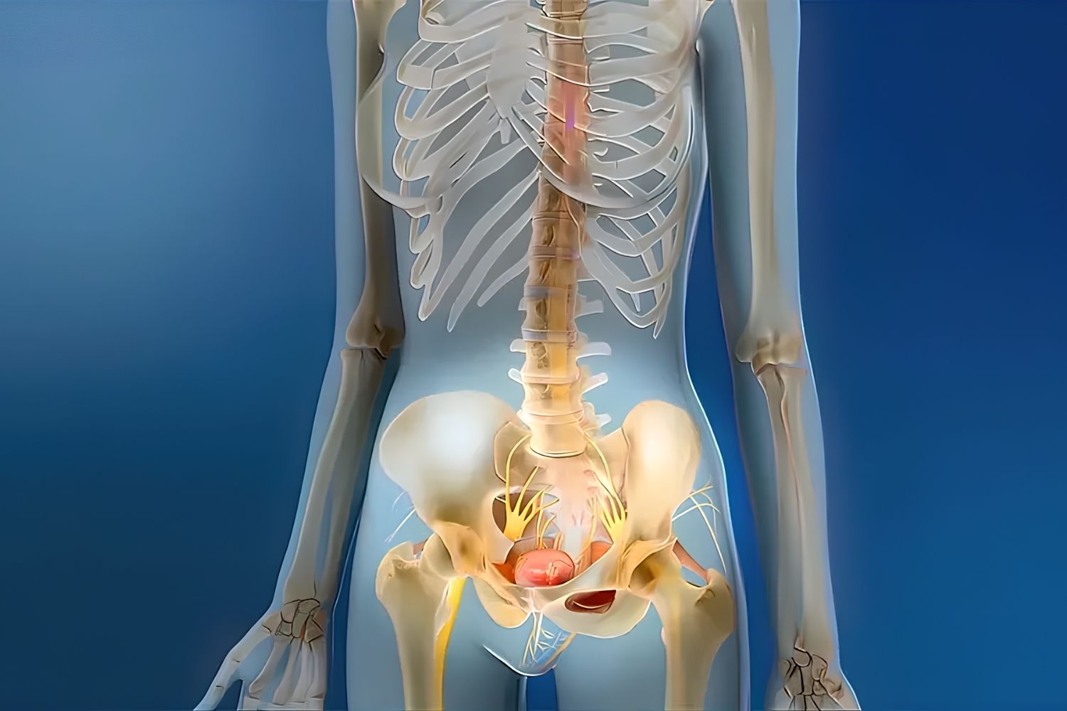
Martin-Gruber anastomosis is a fascinating neural connection in the forearm that not many people know about. This unique anatomical feature involves a nerve crossover between the median and ulnar nerves. Why is it important? Because it can affect how doctors diagnose and treat nerve injuries in the arm. Imagine having a nerve injury and the usual tests don't quite add up—Martin-Gruber anastomosis could be the reason. Understanding this connection helps in avoiding misdiagnosis and ensuring proper treatment. Let's dive into 30 intriguing facts about this lesser-known but crucial anatomical feature.
Key Takeaways:
- Martin-Gruber Anastomosis is a unique nerve connection in the forearm that can affect hand function and sensation in 15-30% of people. Understanding its types and clinical significance is crucial for accurate diagnosis and treatment.
- Detecting Martin-Gruber Anastomosis involves nerve studies, EMG tests, ultrasound, and MRI scans. It can mimic nerve injuries, affect surgeries, and influence carpal tunnel syndrome diagnosis. Ongoing research aims to uncover more about its genetic factors and impact on nerve regeneration.
What is Martin-Gruber Anastomosis?
Martin-Gruber Anastomosis (MGA) is a fascinating neural connection in the human body. It involves a nerve crossover between the median and ulnar nerves in the forearm. This unique connection can affect muscle function and sensation in the hand.
-
MGA is named after two anatomists: The connection is named after Friedrich Martin and Wilhelm Gruber, who first described it in the 19th century.
-
Occurs in about 15-30% of people: Not everyone has this neural connection. Studies show that it appears in roughly 15-30% of the population.
-
Involves median and ulnar nerves: MGA specifically involves a crossover between the median nerve and the ulnar nerve in the forearm.
-
Can affect hand function: This crossover can influence muscle control and sensation in the hand, sometimes causing unexpected clinical symptoms.
Types of Martin-Gruber Anastomosis
There are different types of MGA, categorized based on the specific nerve fibers involved and their pathways. Understanding these types helps in diagnosing and treating related conditions.
-
Type I MGA: Involves motor fibers from the median nerve crossing to the ulnar nerve.
-
Type II MGA: Sensory fibers from the median nerve cross to the ulnar nerve.
-
Type III MGA: Both motor and sensory fibers from the median nerve cross to the ulnar nerve.
-
Type IV MGA: Motor fibers from the ulnar nerve cross to the median nerve.
-
Type V MGA: Sensory fibers from the ulnar nerve cross to the median nerve.
Clinical Significance of Martin-Gruber Anastomosis
MGA can have various clinical implications, especially in diagnosing nerve injuries and conditions. Recognizing MGA is crucial for accurate diagnosis and treatment.
-
Can mimic nerve injuries: MGA can sometimes mimic nerve injuries, leading to misdiagnosis.
-
Affects electromyography (EMG) results: The presence of MGA can alter EMG results, which are used to diagnose nerve and muscle disorders.
-
Important in nerve surgeries: Surgeons need to be aware of MGA during nerve surgeries to avoid complications.
-
Influences carpal tunnel syndrome diagnosis: MGA can affect the diagnosis and treatment of carpal tunnel syndrome.
How is Martin-Gruber Anastomosis Detected?
Detecting MGA involves various diagnostic techniques, including physical examinations and advanced imaging methods. Accurate detection is essential for proper medical management.
-
Nerve conduction studies: These studies measure how well nerves can send electrical signals, helping to detect MGA.
-
Electromyography (EMG): EMG tests the electrical activity of muscles and can reveal the presence of MGA.
-
Ultrasound imaging: High-resolution ultrasound can visualize the nerve pathways and detect MGA.
-
MRI scans: Magnetic Resonance Imaging (MRI) can provide detailed images of nerve structures, aiding in the detection of MGA.
Interesting Facts About Martin-Gruber Anastomosis
MGA is not just a medical curiosity; it has some intriguing aspects that make it a subject of interest for both medical professionals and anatomy enthusiasts.
-
First described in the 19th century: MGA was first documented by Friedrich Martin in 1763 and later by Wilhelm Gruber in 1870.
-
More common in males: Studies suggest that MGA is slightly more common in males than females.
-
Can be bilateral: In some cases, MGA can occur in both forearms.
-
Varies among populations: The prevalence of MGA can vary among different ethnic and population groups.
-
Can affect muscle strength: MGA can influence the strength of certain hand muscles, affecting grip and dexterity.
Challenges in Studying Martin-Gruber Anastomosis
Researching MGA presents unique challenges due to its variability and the complexity of nerve anatomy. Overcoming these challenges is essential for advancing our understanding of this neural connection.
-
Variability in occurrence: The occurrence of MGA varies widely, making it challenging to study.
-
Complex nerve pathways: The intricate pathways of the median and ulnar nerves add to the complexity of studying MGA.
-
Limited awareness: Many medical professionals may not be fully aware of MGA, leading to underdiagnosis.
-
Need for advanced technology: Detecting MGA often requires advanced diagnostic tools, which may not be readily available in all medical settings.
Future Research on Martin-Gruber Anastomosis
Ongoing research aims to uncover more about MGA, its implications, and potential treatments. Future studies will likely focus on improving diagnostic techniques and understanding the genetic factors involved.
-
Genetic factors: Researchers are exploring the genetic factors that may influence the occurrence of MGA.
-
Improving diagnostic methods: Advances in imaging and diagnostic techniques will help in better detecting and understanding MGA.
-
Impact on nerve regeneration: Studying MGA can provide insights into nerve regeneration and repair, benefiting patients with nerve injuries.
-
Educational initiatives: Increasing awareness and education about MGA among medical professionals will improve diagnosis and treatment outcomes.
The Fascinating World of Martin-Gruber Anastomosis
Martin-Gruber Anastomosis (MGA) is a unique nerve connection in the forearm. This connection between the median and ulnar nerves can affect hand movements and sensations. Understanding MGA helps in diagnosing nerve injuries and planning surgeries.
MGA varies among individuals. Some have a strong connection, while others have a weak or absent one. This variation can influence how symptoms present in nerve injuries or conditions like carpal tunnel syndrome.
Knowing about MGA is crucial for healthcare professionals. It aids in accurate diagnosis and effective treatment. For patients, understanding this can explain unusual symptoms and guide them in seeking appropriate care.
In short, Martin-Gruber Anastomosis is a small but significant detail in the complex network of our nervous system. It highlights the incredible variability and adaptability of the human body.
Frequently Asked Questions
Was this page helpful?
Our commitment to delivering trustworthy and engaging content is at the heart of what we do. Each fact on our site is contributed by real users like you, bringing a wealth of diverse insights and information. To ensure the highest standards of accuracy and reliability, our dedicated editors meticulously review each submission. This process guarantees that the facts we share are not only fascinating but also credible. Trust in our commitment to quality and authenticity as you explore and learn with us.
