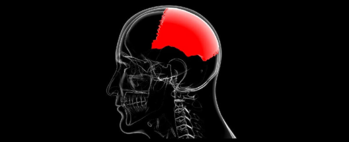
The human body is truly a remarkable creation, with intricate systems and structures that continue to captivate the imagination of both scientists and ordinary individuals alike. Among the numerous bones that make up the skeleton, the parietal bones are a pair that contribute significantly to the overall structure and protection of the brain.
In this article, we will delve into the world of parietal bones and uncover 20 unbelievable facts about them. From their anatomical features and functions to their unique characteristics and variations, you will gain a deeper understanding of these fascinating bones that play a vital role in our daily lives.
So, let’s embark on this journey of discovery and explore the hidden wonders of the parietal bones that lie beneath the surface of our skulls.
Key Takeaways:
- Parietal bones protect the brain and help us feel touch, pain, and temperature. They also play a role in spatial awareness and maintaining skull shape.
- These bones are strong, resistant to fractures, and have unique blood supply. They can vary in size and shape, and even have individual patterns like fingerprints.
The parietal bones are paired symmetrical bones.
Located on the sides and top of the head, each individual has two parietal bones that join together at the midline of the skull.
The parietal bones form the majority of the skull’s roof.
These bones contribute to the formation of the cranium and help provide crucial protection for the brain.
The parietal bones have distinct borders.
These bones are bounded by several other bones, including the frontal, occipital, temporal, and sphenoid bones.
The parietal bones are essential for sensory perception.
They house the parietal lobes of the brain, which are responsible for processing sensory information, including touch, pain, pressure, and temperature.
The parietal bones play a role in spatial awareness.
They are involved in perception of the body’s position in space and assist in orienting movements.
The parietal bones can vary in size and shape.
Individuals may have slight variations in the size and shape of their parietal bones, though they generally maintain a consistent structure.
The parietal bones undergo changes throughout life.
During childhood, the parietal bones are not fully fused and continue to develop and grow. They eventually fuse together by adulthood.
The parietal bones are strong and resistant to fractures.
Due to their thick structure, the parietal bones are highly durable and can withstand significant impacts without breaking.
The parietal bones have a unique blood supply.
They receive blood from the middle meningeal artery, which runs through a groove on the inner surface of the bones.
The parietal bones contribute to the formation of the sagittal suture.
The sagittal suture is a fibrous joint between the two parietal bones, extending along the midline of the skull.
The parietal bones provide attachment points for muscles and ligaments.
Various muscles and ligaments, including those involved in jaw movement and neck support, attach to the parietal bones.
The parietal bones can display individual variations.
Just like fingerprints, the patterns on the inner surface of the parietal bones can be unique to each individual.
The parietal bones are crucial for maintaining skull shape.
They contribute to the overall structure and formation of the skull, helping to maintain its shape and integrity.
The parietal bones contain diploic veins.
These veins run through the inner part of the parietal bones, aiding in the drainage of blood from the skull.
The parietal bones house the superior sagittal sinus.
This sinus is a large blood channel that runs along the midline of the skull and assists in draining blood from the brain.
The parietal bones develop from ossification centers.
During fetal development, the parietal bones form from different ossification centers and gradually merge together.
The parietal bones show age-related changes.
As individuals age, the parietal bones may become thinner and exhibit decreased bone density.
The parietal bones have connections to the temporal and occipital bones.
These connections form important cranial sutures that contribute to the overall strength and stability of the skull.
The parietal bones contribute to the formation of the cerebral falx.
The cerebral falx is a tough, crescent-shaped membrane that separates the two cerebral hemispheres and attaches to the inner surface of the parietal bones.
The parietal bones can undergo surgical procedures.
In certain medical conditions or during surgical interventions, the parietal bones can be manipulated or undergo surgical procedures for therapeutic purposes.
The parietal bones are remarkable structures that play a vital role in protecting the brain and facilitating sensory perception. Understanding these 20 unbelievable facts about parietal bones provides a glimpse into the complexity and significance of these bones within the human skull.
Conclusion
The parietal bones are truly fascinating structures that play a crucial role in protecting our brain and supporting various functions. From their unique shape to their intricate connections with other skull bones, there is so much to discover about these incredible bones. By understanding the anatomy and functions of the parietal bones, we gain insight into the complexity of the human body and the importance of its interdependent systems. Whether you’re a medical professional or simply curious about the human anatomy, exploring the unbelievable facts about the parietal bones offers a deeper appreciation for the wonders of our own bodies.
FAQs
1. What is the function of the parietal bones?
The parietal bones provide protection for the brain and help form the sides and roof of the cranial cavity. They also contribute to the formation of the skull and play a role in sensory perception.
2. How many parietal bones are there in the human skull?
There are two parietal bones in the human skull, one on each side. They are located towards the middle of the skull and are paired with other bones, such as the frontal bone and the occipital bone.
3. Are the parietal bones connected to other bones?
Yes, the parietal bones are connected to several other bones in the skull. They articulate with the frontal bone in the front, the occipital bone at the back, and the temporal bones on the sides.
4. Can the parietal bones be affected by injuries or diseases?
Yes, like other bones in the body, the parietal bones can be subject to injuries such as fractures or trauma. Certain diseases or conditions, such as craniosynostosis, can also affect the development or shape of the parietal bones.
5. Can the parietal bones regenerate if they are damaged?
No, the parietal bones do not have the ability to regenerate if they are damaged. However, with proper medical care and treatment, fractures or injuries to the parietal bones can heal over time.
Was this page helpful?
Our commitment to delivering trustworthy and engaging content is at the heart of what we do. Each fact on our site is contributed by real users like you, bringing a wealth of diverse insights and information. To ensure the highest standards of accuracy and reliability, our dedicated editors meticulously review each submission. This process guarantees that the facts we share are not only fascinating but also credible. Trust in our commitment to quality and authenticity as you explore and learn with us.
