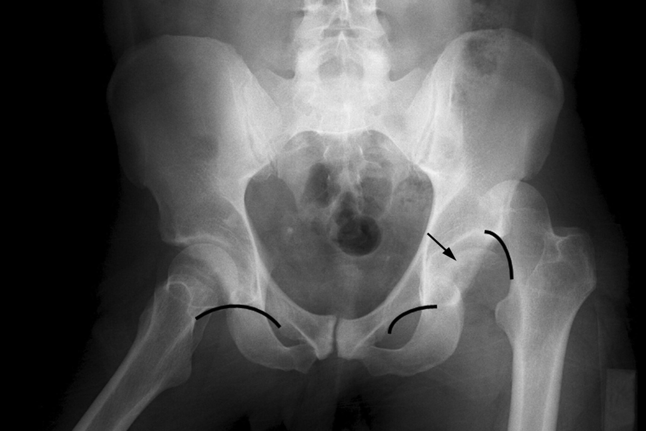
Pipkin fracture-dislocation might sound like a mouthful, but understanding it can be straightforward. This injury involves a break in the femoral head, often accompanied by a hip dislocation. Commonly caused by high-impact trauma, such as car accidents or falls, it requires immediate medical attention. Symptoms include severe hip pain, inability to move the leg, and visible deformity. Treatment options range from surgical intervention to physical therapy, depending on the severity. Knowing the facts about Pipkin fracture-dislocation can help you recognize the signs and understand the treatment process. Let's dive into 30 crucial facts about this complex injury.
Key Takeaways:
- Pipkin fracture-dislocation is a rare and severe hip injury named after Dr. Pipkin. It requires immediate medical attention and can lead to complications like avascular necrosis and arthritis.
- Understanding the types, symptoms, and treatment options for Pipkin fractures is crucial for effective recovery. Younger patients are more susceptible to this injury due to high-energy trauma.
What is a Pipkin Fracture-Dislocation?
A Pipkin fracture-dislocation is a specific type of injury involving the hip joint. It occurs when the femoral head (the ball of the hip joint) fractures and dislocates from the acetabulum (the socket of the hip joint). This injury is often severe and requires immediate medical attention.
-
Named After Dr. Pipkin: The term "Pipkin fracture" is named after Dr. Garry Pipkin, who first described this type of injury in 1957.
-
Femoral Head Involvement: This fracture specifically involves the femoral head, which is the rounded top part of the thigh bone that fits into the hip socket.
-
Common in High-Energy Trauma: Pipkin fractures typically result from high-energy trauma, such as car accidents or falls from significant heights.
-
Classification System: Pipkin fractures are classified into four types based on the location and extent of the fracture.
Types of Pipkin Fractures
Understanding the different types of Pipkin fractures helps in determining the appropriate treatment and prognosis.
-
Type I: Involves a fracture of the femoral head below the fovea capitis, which does not affect the weight-bearing surface.
-
Type II: Involves a fracture above the fovea capitis, affecting the weight-bearing surface of the femoral head.
-
Type III: Combines a Type I or II fracture with a fracture of the femoral neck.
-
Type IV: Involves a Type I or II fracture along with a fracture of the acetabulum.
Symptoms and Diagnosis
Recognizing the symptoms and understanding the diagnostic process is crucial for timely and effective treatment.
-
Severe Hip Pain: Patients typically experience intense pain in the hip region, making it difficult to move the leg.
-
Visible Deformity: The affected leg may appear shorter and rotated compared to the uninjured leg.
-
Limited Mobility: Movement of the hip joint is usually severely restricted due to pain and mechanical blockage.
-
X-Rays: Initial diagnosis often involves X-rays to visualize the fracture and dislocation.
-
CT Scans: Computed tomography (CT) scans provide detailed images, helping to assess the extent of the injury.
Treatment Options
Treatment for Pipkin fractures varies based on the type and severity of the injury.
-
Closed Reduction: In some cases, the dislocated femoral head can be repositioned without surgery through a process called closed reduction.
-
Open Reduction and Internal Fixation (ORIF): Surgery involving the repositioning of the bone and securing it with screws or plates is often necessary.
-
Hip Replacement: Severe cases may require partial or total hip replacement, especially in older patients.
-
Physical Therapy: Post-surgery, physical therapy is crucial for regaining strength and mobility in the hip joint.
Complications and Prognosis
Understanding potential complications and the overall prognosis helps in managing expectations and planning recovery.
-
Avascular Necrosis: One of the most serious complications is avascular necrosis, where the blood supply to the femoral head is disrupted, leading to bone death.
-
Post-Traumatic Arthritis: Patients may develop arthritis in the hip joint due to the injury and subsequent changes in joint mechanics.
-
Infection: Surgical treatment carries a risk of infection, which can complicate recovery.
-
Recurrent Dislocation: There is a risk of the hip dislocating again, especially if the initial injury was severe.
-
Delayed Union or Nonunion: Sometimes, the fractured bone may heal slowly or not at all, requiring additional interventions.
Recovery and Rehabilitation
Recovery from a Pipkin fracture-dislocation involves a combination of medical treatment, physical therapy, and lifestyle adjustments.
-
Weight-Bearing Restrictions: Patients are often advised to avoid putting weight on the injured leg for several weeks to allow proper healing.
-
Pain Management: Medications and other pain management strategies are essential during the initial recovery phase.
-
Gradual Increase in Activity: Physical activity is gradually increased under the supervision of a healthcare provider to ensure safe and effective rehabilitation.
-
Use of Assistive Devices: Crutches or walkers may be necessary to aid mobility during the recovery period.
-
Long-Term Follow-Up: Regular follow-up appointments are crucial to monitor healing and address any complications promptly.
Interesting Facts About Pipkin Fracture-Dislocation
Here are some additional intriguing facts about this specific type of injury.
-
Rare Injury: Pipkin fractures are relatively rare compared to other types of hip fractures and dislocations.
-
Younger Patients: This injury is more common in younger patients due to the high-energy trauma typically required to cause it.
-
Historical Cases: Historical records show that similar injuries were treated even before modern medical techniques, though with much less success.
Final Thoughts on Pipkin Fracture-Dislocation
Pipkin fracture-dislocation, a complex injury involving the femoral head and hip joint, demands immediate medical attention. Understanding the types of Pipkin fractures can help in recognizing the severity and necessary treatment. Type I involves a fracture below the fovea, while Type II includes a fracture above the fovea. Type III combines a Type I or II fracture with a femoral neck fracture, and Type IV adds an acetabular fracture to the mix.
Treatment options range from non-surgical methods like traction and bed rest to surgical interventions such as open reduction and internal fixation. Early diagnosis and appropriate management are crucial for preventing complications like avascular necrosis and post-traumatic arthritis.
Being aware of these facts can aid in better understanding and managing this serious injury. Always consult a healthcare professional for accurate diagnosis and treatment.
Frequently Asked Questions
Was this page helpful?
Our commitment to delivering trustworthy and engaging content is at the heart of what we do. Each fact on our site is contributed by real users like you, bringing a wealth of diverse insights and information. To ensure the highest standards of accuracy and reliability, our dedicated editors meticulously review each submission. This process guarantees that the facts we share are not only fascinating but also credible. Trust in our commitment to quality and authenticity as you explore and learn with us.
