


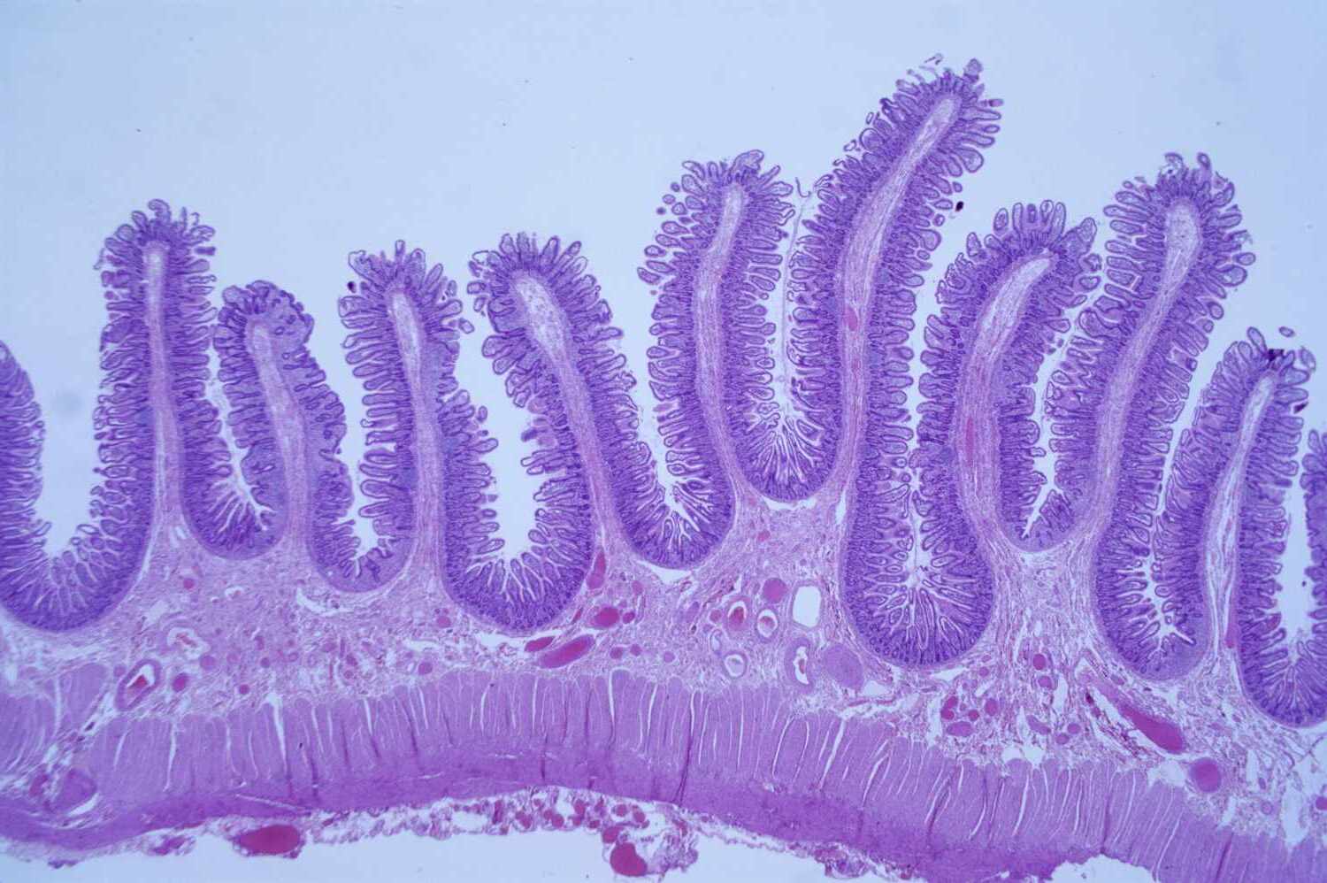
Histology is the study of tissues at the microscopic level. Ever wondered what makes up the organs in your body? Histology holds the answers. This field helps scientists and doctors understand how tissues function, how diseases affect them, and how they can be treated. From examining the tiny cells in your liver to understanding the complex structures in your brain, histology covers it all. Microscopes play a crucial role, allowing us to see what the naked eye cannot. Whether you're a student, a curious mind, or someone in the medical field, these 30 facts about histology will give you a deeper appreciation for the intricate world of tissues. Ready to dive in? Let's get started!
Key Takeaways:
- Histology, the study of tissues, uses microscopes and special dyes to understand how diseases affect organisms. It's crucial in diagnosing cancer and developing new treatments.
- Histology has diverse applications, from botany to criminal investigations. It helps diagnose diseases, study developmental biology, and assess the impact of environmental toxins on health.
What is Histology?
Histology, the study of tissues, reveals the microscopic structure of plants and animals. It plays a crucial role in understanding how organisms function and how diseases affect them. Here are some fascinating facts about histology.
-
Histology comes from the Greek words "histos" meaning tissue and "logia" meaning study. This field focuses on examining tissues under a microscope.
-
Histologists use special dyes to stain tissues, making different structures visible. Hematoxylin and eosin (H&E) staining is the most common technique.
-
Histology helps diagnose diseases. Pathologists examine tissue samples to identify abnormalities like cancer.
-
The first histology textbook was written by Marie François Xavier Bichat in 1801. He is often called the father of modern histology.
-
Histology is essential in medical research. It helps scientists understand how diseases progress and develop new treatments.
Tools and Techniques in Histology
Histologists use various tools and techniques to study tissues. These methods have evolved over time, improving our understanding of microscopic structures.
-
Microscopes are the primary tool in histology. Light microscopes, electron microscopes, and confocal microscopes each offer different levels of detail.
-
Tissue samples are often embedded in paraffin wax. This process makes them easier to slice into thin sections for examination.
-
Microtomes are used to cut tissue samples into thin sections. These slices are typically only a few micrometers thick.
-
Immunohistochemistry (IHC) is a technique that uses antibodies to detect specific proteins in tissues. This method helps identify diseases like cancer.
-
Fluorescence microscopy uses fluorescent dyes to label specific structures within tissues. This technique allows scientists to study the distribution of molecules.
Histology in Different Fields
Histology isn't just for medical research. It has applications in various fields, from botany to forensic science.
-
Plant histology studies the tissues of plants. It helps botanists understand plant structure and function.
-
Veterinary histology examines tissues from animals. This field helps diagnose diseases in pets and livestock.
-
Forensic histology aids in criminal investigations. Tissue samples can provide clues about the cause of death or the presence of toxins.
-
Histology is crucial in developmental biology. It helps scientists study how tissues and organs form during embryonic development.
-
Environmental histology examines the effects of pollutants on tissues. This field helps assess the impact of environmental toxins on health.
Histology and Disease
Understanding tissues at the microscopic level is vital for diagnosing and treating diseases. Histology provides insights into how diseases affect the body.
-
Cancer diagnosis often relies on histology. Pathologists examine tissue samples to identify cancerous cells and determine the type and stage of cancer.
-
Histology helps diagnose infectious diseases. Tissue samples can reveal the presence of bacteria, viruses, or fungi.
-
Autoimmune diseases can be studied through histology. Tissue samples show how the immune system attacks the body's own tissues.
-
Histology is used to study neurodegenerative diseases. Examining brain tissues helps researchers understand conditions like Alzheimer's and Parkinson's.
-
Liver diseases can be diagnosed with histology. Tissue samples reveal damage caused by conditions like hepatitis or cirrhosis.
The Future of Histology
Histology continues to evolve with new technologies and techniques. These advancements promise to improve our understanding of tissues and diseases.
-
Digital histology uses computer technology to analyze tissue samples. This method allows for more precise and efficient diagnosis.
-
Artificial intelligence (AI) is being integrated into histology. AI can help identify patterns in tissue samples that might be missed by human eyes.
-
3D histology provides a more comprehensive view of tissues. This technique reconstructs tissues in three dimensions, offering new insights into their structure.
-
Single-cell histology studies individual cells within tissues. This method helps researchers understand cellular diversity and function.
-
Advances in staining techniques are improving histology. New dyes and markers allow for more detailed and specific labeling of tissue structures.
Interesting Historical Facts
Histology has a rich history filled with intriguing discoveries and milestones. These historical facts highlight the development of this fascinating field.
-
The invention of the microscope in the 17th century revolutionized histology. Antonie van Leeuwenhoek, a Dutch scientist, was one of the first to observe cells under a microscope.
-
Marcello Malpighi, an Italian biologist, is considered the founder of microscopic anatomy. He made significant contributions to the study of tissues in the 17th century.
-
In the 19th century, Rudolf Virchow, a German physician, advanced the field of histology. He proposed that diseases originate at the cellular level.
-
The discovery of the Golgi apparatus in 1898 by Camillo Golgi was a major milestone. This cellular structure plays a crucial role in processing and packaging proteins.
-
The development of electron microscopy in the 20th century allowed for unprecedented detail in tissue examination. This technology revealed structures previously invisible with light microscopes.
The Microscopic World Awaits
Histology, the study of tissues, offers a fascinating glimpse into the microscopic world that makes up living organisms. From understanding how tissues function to diagnosing diseases, histology plays a crucial role in medicine and research. Knowing these 30 facts can deepen your appreciation for this intricate field.
Whether you're a student, a professional, or just curious, histology provides valuable insights into the complexity of life. It helps us understand how our bodies work and how we can keep them healthy. So next time you look at a microscope slide, remember the incredible details and stories each tissue holds.
Keep exploring, keep learning, and let your curiosity guide you through the wonders of histology. The microscopic world is full of surprises, and there's always more to discover. Happy studying!
Frequently Asked Questions
Was this page helpful?
Our commitment to delivering trustworthy and engaging content is at the heart of what we do. Each fact on our site is contributed by real users like you, bringing a wealth of diverse insights and information. To ensure the highest standards of accuracy and reliability, our dedicated editors meticulously review each submission. This process guarantees that the facts we share are not only fascinating but also credible. Trust in our commitment to quality and authenticity as you explore and learn with us.