

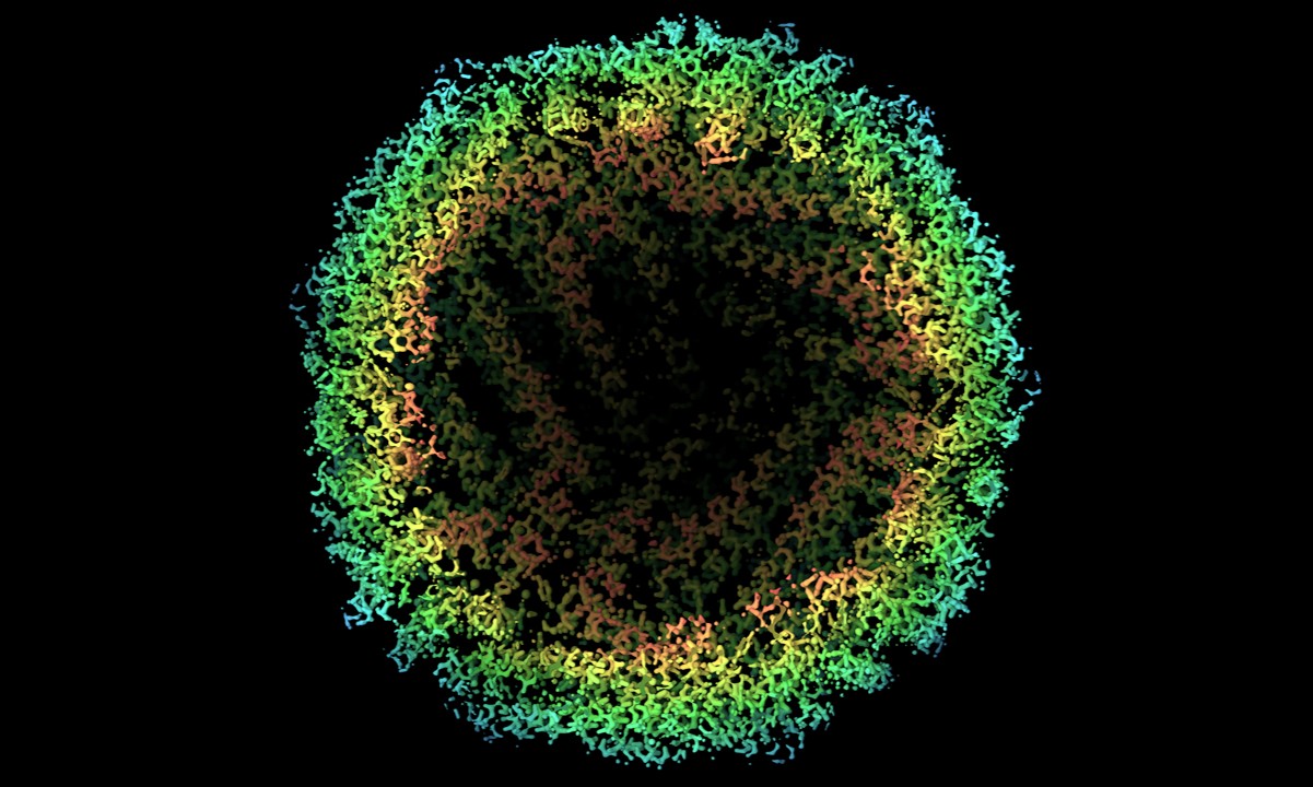
Cryo-electron microscopy (cryo-EM) is a groundbreaking technique revolutionizing the field of structural biology. Ever wondered how scientists can visualize the tiniest structures within cells? Cryo-EM allows researchers to capture images of proteins, viruses, and other macromolecules in their natural state, frozen at cryogenic temperatures. This method provides unprecedented detail, helping to unravel the mysteries of molecular machinery. Unlike traditional microscopy, which can damage delicate samples, cryo-EM preserves their integrity, offering a clearer view. From aiding drug discovery to understanding disease mechanisms, this technology is a game-changer. Ready to dive into the fascinating world of cryo-electron microscopy? Let's explore 29 amazing facts that highlight its significance and impact.
What is Cryo-electron Microscopy?
Cryo-electron microscopy (Cryo-EM) is a cutting-edge technique used to observe the fine details of biological molecules. This method involves freezing samples at extremely low temperatures to preserve their natural structure.
- Cryo-EM allows scientists to see proteins and other molecules in their natural state without the need for dyes or stains.
- This technique can capture images of molecules at near-atomic resolution, providing incredible detail.
- Cryo-EM has revolutionized structural biology by allowing researchers to study the shapes and functions of complex molecules.
- The method involves rapidly freezing samples in liquid ethane to prevent ice crystal formation, which can damage the sample.
- Cryo-EM uses electron beams instead of light to create images, which allows for much higher resolution.
The History of Cryo-electron Microscopy
Understanding the history of Cryo-EM helps appreciate its significance in modern science. From its inception to its current state, Cryo-EM has undergone significant advancements.
- The concept of electron microscopy dates back to the 1930s, but Cryo-EM was developed in the 1980s.
- Jacques Dubochet, Joachim Frank, and Richard Henderson were awarded the Nobel Prize in Chemistry in 2017 for their work in developing Cryo-EM.
- Early electron microscopes required samples to be dehydrated and stained, which could alter their natural structure.
- The development of vitrification, a process of rapid freezing, was a game-changer for Cryo-EM.
- Advances in detector technology and image processing software have significantly improved the resolution and speed of Cryo-EM.
How Cryo-electron Microscopy Works
The process of Cryo-EM involves several steps, each crucial for obtaining high-quality images. Here's a breakdown of how it works.
- Samples are first prepared by placing them on a grid and then rapidly freezing them in liquid ethane.
- The frozen samples are then transferred to the electron microscope, which operates at cryogenic temperatures.
- An electron beam is directed at the sample, and the electrons that pass through are captured to form an image.
- Multiple images are taken from different angles to create a 3D reconstruction of the molecule.
- Advanced software is used to process and combine these images, resulting in a detailed 3D model.
Applications of Cryo-electron Microscopy
Cryo-EM has a wide range of applications in various fields of science and medicine. Its ability to provide detailed images of biological molecules makes it invaluable.
- Cryo-EM is used to study the structure of viruses, helping in the development of vaccines and antiviral drugs.
- Researchers use Cryo-EM to understand the mechanisms of enzymes and other proteins, which can lead to new drug discoveries.
- This technique has been instrumental in studying the structure of ribosomes, the molecular machines that make proteins in cells.
- Cryo-EM helps in understanding the structure of membrane proteins, which are targets for many drugs.
- It has also been used to study large molecular complexes, such as those involved in DNA replication and repair.
Advantages of Cryo-electron Microscopy
Cryo-EM offers several advantages over traditional microscopy techniques, making it a preferred choice for many researchers.
- Unlike X-ray crystallography, Cryo-EM does not require the crystallization of samples, which can be difficult for some molecules.
- Cryo-EM can be used to study large and complex molecules that are challenging to analyze with other techniques.
- The method preserves the natural state of the sample, providing more accurate structural information.
- Cryo-EM allows for the study of dynamic processes by capturing images of molecules in different states.
- The technique is less damaging to samples compared to other electron microscopy methods, as it uses lower doses of electrons.
Challenges and Future of Cryo-electron Microscopy
Despite its advantages, Cryo-EM also faces some challenges. However, ongoing research and technological advancements continue to push the boundaries of what this technique can achieve.
- One of the main challenges is the need for expensive and sophisticated equipment, which can be a barrier for some research institutions.
- Preparing high-quality samples for Cryo-EM can be technically demanding and time-consuming.
- The technique requires skilled operators and extensive training to achieve optimal results.
- Future developments in detector technology, image processing algorithms, and sample preparation methods are expected to further enhance the capabilities of Cryo-EM.
The Power of Cryo-electron Microscopy
Cryo-electron microscopy (cryo-EM) has revolutionized how scientists view biological molecules. This technique allows researchers to see proteins and viruses in their natural state, frozen in time. By avoiding the need for dyes or stains, cryo-EM provides a clearer, more accurate picture of molecular structures. This has led to breakthroughs in understanding diseases and developing new treatments.
Cryo-EM's ability to capture high-resolution images has made it a game-changer in structural biology. Scientists can now study complex molecules that were once too difficult to analyze. This has opened doors to new discoveries and innovations in medicine and biology.
The future of cryo-EM looks promising, with advancements in technology making it even more powerful. As researchers continue to push the boundaries, cryo-EM will undoubtedly play a crucial role in scientific discoveries for years to come.
Was this page helpful?
Our commitment to delivering trustworthy and engaging content is at the heart of what we do. Each fact on our site is contributed by real users like you, bringing a wealth of diverse insights and information. To ensure the highest standards of accuracy and reliability, our dedicated editors meticulously review each submission. This process guarantees that the facts we share are not only fascinating but also credible. Trust in our commitment to quality and authenticity as you explore and learn with us.