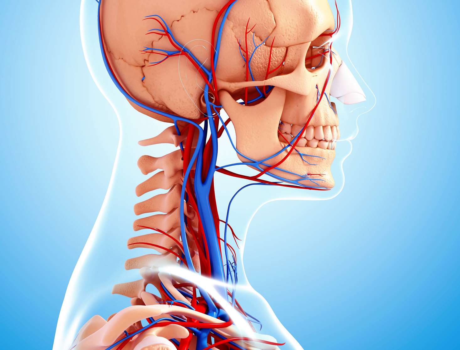
The external jugular vein is an essential part of the human anatomy that plays a crucial role in the transportation of blood. Situated in the neck region, this vein is responsible for draining deoxygenated blood from the head, face, and neck back to the heart. The external jugular vein runs superficially along the side of the neck, making it easily accessible for medical procedures and examinations.
Despite its significance, many people are unaware of the astounding facts surrounding the external jugular vein. In this article, we will explore 18 fascinating and little-known facts about the external jugular vein. From its anatomy and function to its significance in medical procedures, these facts will deepen your understanding of the human body and its intricate mechanisms.
Key Takeaways:
- The external jugular vein is a vital blood vessel in the neck that helps circulate deoxygenated blood back to the heart, and its visibility makes it useful for medical procedures like drawing blood.
- Postural changes can affect the blood flow in the external jugular vein, and its pulsations provide valuable information about a patient’s condition in emergency medicine.
The Location of the External Jugular Vein
The external jugular vein can be found in the neck, running diagonally over the surface of the sternocleidomastoid muscle. It is easily visible and is often used for medical procedures such as drawing blood or administering intravenous medications.
The Function of the External Jugular Vein
The primary function of the external jugular vein is to collect deoxygenated blood from the superficial tissues of the head, face, and neck. It then transports this blood back to the heart, where it can be reoxygenated and circulated to the rest of the body.
The Size of the External Jugular Vein
The external jugular vein varies in size, ranging from about 0.5 to 1.5 centimeters in diameter. Its size may increase or decrease depending on factors such as hydration levels, physical activity, and overall health.
Connectivity with the Superior Vena Cava
The external jugular vein connects with the subclavian vein to form the brachiocephalic vein, which then merges with the other brachiocephalic vein to form the superior vena cava. This major vein is responsible for returning blood from the upper body back to the heart.
Valve Structure
The external jugular vein contains numerous valves that help regulate blood flow. These valves prevent backflow and ensure that blood moves in the correct direction, towards the heart.
Collateral Circulation
The external jugular vein is part of a complex network of blood vessels known as collateral circulation. In instances where the main blood vessels are obstructed or damaged, collateral circulation ensures that blood can still reach its destination using alternative pathways.
Pulsation of the External Jugular Vein
The external jugular vein exhibits a distinct pulsation, which can be observed visually or palpated. This pulsation is synchronized with the heartbeat and reflects the blood flow through the vein.
Accessibility for Medical Procedures
The external jugular vein is easily accessible for medical procedures such as phlebotomy, where blood is drawn for diagnostic tests. Its visibility and location make it a preferred choice for healthcare professionals.
Prone to Distention
The external jugular vein has a tendency to become distended or swollen. This can occur due to factors like physical exertion, changes in body position, or increased pressure within the vein itself.
Role in Circulation and Drainage
The external jugular vein serves as an important pathway for both blood circulation and lymphatic drainage in the head and neck region. It ensures the proper flow of fluids, nutrients, and waste materials throughout the body.
Possible Sites for Catheter Placement
Due to its accessibility and size, the external jugular vein is often considered for central venous catheter placement. This procedure involves inserting a catheter into a large vein to administer medications, fluids, or nutrition.
Variations in Anatomy
The anatomy of the external jugular vein can vary from person to person. While the general location and course remain consistent, there may be differences in size or branching patterns.
Pressure Measurement
The external jugular vein can be used to measure the central venous pressure, which reflects the pressure in the right atrium of the heart. This measurement provides valuable information about the heart’s function and overall fluid balance in the body.
Connection to the Cervical Plexus
The external jugular vein is closely associated with the cervical plexus, a network of nerves that supplies sensation to the neck and head. These nerves run alongside and often intertwine with the vein.
Used for Localization in Surgical Procedures
In certain surgical procedures, surgeons may use the external jugular vein as a landmark to locate and identify specific structures in the neck and surrounding areas. Its visibility aids in precise surgical navigation.
Effects of Postural Changes
Postural changes, such as standing up or lying down, can impact the blood flow within the external jugular vein. The position of the body affects the extent of distension or collapse of the vein.
Potential for Complications
Like any major blood vessel, the external jugular vein carries a risk of complications. These can include thrombosis (blood clot formation), infection, or injury during medical procedures.
Importance in Emergency Medicine
In emergency medicine, the external jugular vein is often used as a reference point for assessing a patient’s condition. Its visibility and pulsations provide valuable information about blood circulation and fluid status.
These 18 astounding facts about the external jugular vein demonstrate its significance in the human body’s circulatory and lymphatic systems. Understanding its anatomy and functions helps healthcare professionals in various medical procedures and contributes to the overall knowledge of human physiology.
Conclusion
The external jugular vein is a fascinating component of the human anatomy, serving a crucial role in the circulatory system. Its prominent location and unique features make it a subject of interest for medical professionals and curious individuals alike.
Throughout this article, we have learned some astounding facts about the external jugular vein. From its role in draining blood from the head and neck to its connection to the superior vena cava, the knowledge gained about this vein helps us understand the complexity and intricacy of the human body.
As we continue to unravel the mysteries of the human anatomy, the external jugular vein stands out as an important piece of the puzzle. Its functions, structure, and significance remind us of the incredible design and functionality of our bodies.
FAQs
1. What is the external jugular vein?
The external jugular vein is a major blood vessel located in the neck. It receives blood from the scalp, face, and neck regions and drains into the superior vena cava.
2. How does the external jugular vein differ from the internal jugular vein?
The external jugular vein lies on the surface of the neck and is more visible than the internal jugular vein, which is deeper within the neck. The external jugular vein is also responsible for draining superficial structures, while the internal jugular vein plays a role in draining deeper structures of the head and neck.
3. Are there any medical conditions associated with the external jugular vein?
Yes, certain medical conditions can affect the external jugular vein. These include thrombosis (clot formation), compression due to an underlying mass or tumor, and inflammation of the vein known as phlebitis.
4. Can the external jugular vein be used for medical procedures?
Yes, the external jugular vein can be utilized for certain medical procedures, such as blood draws, intravenous medication administration, and central venous access.
5. Can the external jugular vein be easily visualized?
Under normal circumstances, the external jugular vein is relatively easy to locate and visualize, especially when it becomes distended due to increased pressure within the venous system.
Curious about more captivating facts? Uncover the secrets of human anatomy and its enigmatic wonders. Dive into the extraordinary world of the cardiovascular system, where blood flows and hearts beat. Interested in cutting-edge medical education? Explore the fascinating history and groundbreaking research at the University of Texas Southwestern Medical Center. From the depths of our veins to the frontiers of medical science, there's always more to learn and discover. So, keep reading, keep wondering, and never stop marveling at the incredible complexities of the human body and the dedicated professionals who study it.
Was this page helpful?
Our commitment to delivering trustworthy and engaging content is at the heart of what we do. Each fact on our site is contributed by real users like you, bringing a wealth of diverse insights and information. To ensure the highest standards of accuracy and reliability, our dedicated editors meticulously review each submission. This process guarantees that the facts we share are not only fascinating but also credible. Trust in our commitment to quality and authenticity as you explore and learn with us.
