


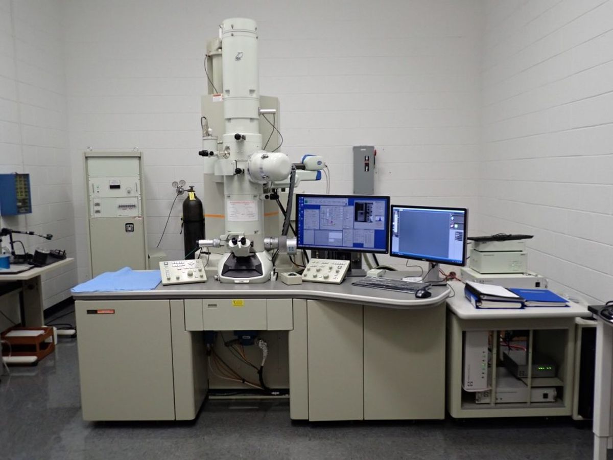
What is an electron microscope? An electron microscope uses a beam of electrons to create an image of a specimen. Unlike light microscopes, which use visible light, electron microscopes can magnify objects up to two million times their actual size. This allows scientists to see incredibly small details, like the structure of cells, viruses, and even individual atoms. There are two main types: transmission electron microscopes (TEM) and scanning electron microscopes (SEM). TEMs pass electrons through a thin sample, while SEMs scan the surface with electrons. Both provide different but complementary views of the microscopic world.
What is an Electron Microscope?
An electron microscope uses a beam of electrons to create an image of a specimen. This technology allows scientists to see objects at a much higher resolution than light microscopes.
-
Electron microscopes can magnify objects up to 10 million times. This level of magnification reveals details at the atomic level.
-
There are two main types: Transmission Electron Microscopes (TEM) and Scanning Electron Microscopes (SEM). TEMs pass electrons through a specimen, while SEMs scan the surface with electrons.
-
The first electron microscope was built in 1931. German engineers Ernst Ruska and Max Knoll created it.
-
Electron microscopes use electromagnetic lenses. These lenses focus the electron beam, similar to how glass lenses focus light in optical microscopes.
-
Specimens must be placed in a vacuum. Air molecules can scatter electrons, so a vacuum is necessary for clear imaging.
How Electron Microscopes Work
Understanding the working principles of electron microscopes can help appreciate their capabilities.
-
Electrons are generated by an electron gun. This device emits electrons that are then accelerated towards the specimen.
-
Specimens are often coated with a thin layer of metal. This coating helps to conduct electrons and produce clearer images.
-
Electrons interact with the specimen to produce an image. These interactions include scattering and absorption.
-
Detectors capture the electrons that pass through or bounce off the specimen. These detectors convert electron signals into an image.
-
Images are displayed on a computer screen. Advanced software processes the electron signals to create detailed images.
Applications of Electron Microscopes
Electron microscopes are used in various fields, from biology to materials science.
-
Biologists use electron microscopes to study cell structures. They can see organelles like mitochondria and ribosomes in great detail.
-
Materials scientists examine the atomic structure of materials. This helps in developing new materials with specific properties.
-
Electron microscopes are crucial in nanotechnology. They allow scientists to manipulate and observe nanoparticles.
-
Forensic scientists use them to analyze evidence. They can examine tiny details on bullets, fibers, and other materials.
-
Geologists study mineral structures. Electron microscopes reveal the composition and formation of minerals.
Advantages of Electron Microscopes
Electron microscopes offer several benefits over traditional light microscopes.
-
Higher resolution and magnification. They can reveal details that are invisible to light microscopes.
-
Ability to see the internal structure of cells. TEMs can show cross-sections of cells and tissues.
-
Detailed surface imaging. SEMs provide 3D images of surfaces.
-
Versatility in applications. They are used in many scientific fields.
-
Advanced imaging techniques. Techniques like cryo-electron microscopy allow for imaging of specimens in their natural state.
Challenges and Limitations
Despite their advantages, electron microscopes have some limitations.
-
High cost. They are expensive to purchase and maintain.
-
Complex operation. Requires specialized training to operate.
-
Specimen preparation can be time-consuming. Preparing specimens for electron microscopy involves several steps.
-
Limited to non-living specimens. The vacuum environment and electron beam can damage living cells.
-
Size and space requirements. They are large and require a dedicated space.
Innovations in Electron Microscopy
Recent advancements have improved the capabilities of electron microscopes.
-
Cryo-electron microscopy has revolutionized structural biology. It allows for imaging of proteins and other biomolecules in their natural state.
-
Development of aberration-corrected electron microscopes. These microscopes correct distortions in the electron beam for clearer images.
-
Integration with other imaging techniques. Combining electron microscopy with techniques like X-ray microscopy provides more comprehensive data.
-
Automated imaging and analysis. Advanced software can automate the imaging process and analyze data more efficiently.
-
Environmental electron microscopy. Allows for imaging of specimens in a more natural state without a high vacuum.
-
In-situ electron microscopy. Enables observation of dynamic processes in real-time, such as chemical reactions and material changes.
The Power of Electron Microscopes
Electron microscopes have revolutionized our understanding of the microscopic world. These powerful tools allow scientists to see details at the atomic level, revealing structures and processes that were once invisible. From medical research to materials science, electron microscopes play a crucial role in advancing technology and knowledge. They help diagnose diseases, develop new materials, and even explore the mysteries of the universe.
Understanding how electron microscopes work and their applications can inspire future innovations. Whether you're a student, a researcher, or just curious, knowing these facts can deepen your appreciation for this incredible technology. So next time you hear about a groundbreaking discovery, remember the electron microscope might have played a part. Keep exploring, keep learning, and who knows what amazing things you'll uncover next!
Was this page helpful?
Our commitment to delivering trustworthy and engaging content is at the heart of what we do. Each fact on our site is contributed by real users like you, bringing a wealth of diverse insights and information. To ensure the highest standards of accuracy and reliability, our dedicated editors meticulously review each submission. This process guarantees that the facts we share are not only fascinating but also credible. Trust in our commitment to quality and authenticity as you explore and learn with us.