


Ever wondered how scientists detect viruses in tissues? The Coons Fluorescent Antibody Method is a groundbreaking technique that uses fluorescent dyes to tag antibodies, making it easier to spot viruses under a microscope. This method, developed by Albert Coons in the 1940s, revolutionized medical research by allowing scientists to see where viruses and bacteria are hiding in the body. Imagine being able to see glowing markers that pinpoint exactly where an infection is! This technique has been crucial in diagnosing diseases, studying immune responses, and even in cancer research. Let's dive into 30 fascinating facts about this incredible method that changed the face of medical science.
Key Takeaways:
- Coons Fluorescent Antibody Method revolutionized medical research by using fluorescent dyes to visualize diseases and cellular processes, aiding in cancer diagnosis and studying brain functions.
- This method has high sensitivity and specificity, allowing real-time observation of living cells, but it requires skilled technicians and proper sample preparation.
What is Coons Fluorescent Antibody Method?
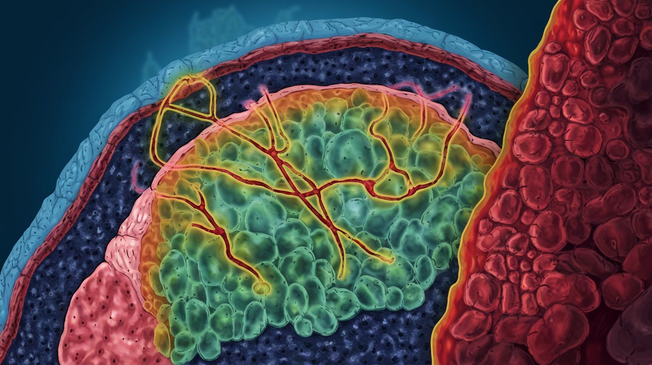
The Coons Fluorescent Antibody Method is a groundbreaking technique in immunology. It allows scientists to visualize specific proteins or antigens in cells and tissues using fluorescent dyes. This method has revolutionized how researchers study diseases and cellular processes.
-
Invented by Albert Coons: Albert Coons developed this method in 1941, making it a cornerstone in immunohistochemistry.
-
Uses Fluorescent Dyes: Fluorescent dyes attach to antibodies, which then bind to specific antigens, illuminating them under a microscope.
-
First Used to Detect Pneumococci: Initially, Coons used this method to detect pneumococci bacteria in infected tissues.
-
Revolutionized Pathology: This technique transformed pathology by allowing precise identification of pathogens in tissue samples.
-
Two Main Types: There are direct and indirect fluorescent antibody methods. Direct uses a single antibody, while indirect uses a secondary antibody for amplification.
How Does It Work?
Understanding the mechanics of the Coons Fluorescent Antibody Method can help grasp its significance. The process involves several steps to ensure accurate results.
-
Antigen-Antibody Binding: The method relies on the specific binding between an antigen and its corresponding antibody.
-
Fluorescent Labeling: Antibodies are tagged with fluorescent dyes, which emit light when exposed to specific wavelengths.
-
Microscopy: A fluorescence microscope is used to visualize the fluorescently labeled antibodies bound to antigens.
-
Sample Preparation: Samples must be carefully prepared and fixed to preserve the structure and antigenicity of the tissues.
-
Blocking Non-Specific Binding: Blocking agents are used to prevent non-specific binding, ensuring only the target antigen is highlighted.
Applications in Medical Research
The Coons Fluorescent Antibody Method has numerous applications in medical research, aiding in the understanding of various diseases.
-
Cancer Research: Used to identify cancer markers in tissues, helping in diagnosis and treatment planning.
-
Infectious Diseases: Detects pathogens like bacteria and viruses in tissue samples, crucial for diagnosing infections.
-
Autoimmune Disorders: Helps identify autoantibodies in autoimmune diseases, providing insights into disease mechanisms.
-
Neuroscience: Used to study the distribution of neurotransmitters and receptors in the brain.
-
Developmental Biology: Assists in studying the expression of proteins during embryonic development.
Advantages of the Method
The Coons Fluorescent Antibody Method offers several advantages over traditional staining techniques.
-
High Sensitivity: Can detect even small amounts of antigen due to the high sensitivity of fluorescent dyes.
-
Specificity: Provides high specificity as antibodies bind only to their target antigens.
-
Quantitative Analysis: Allows for quantitative analysis of antigen levels in tissues.
-
Multiple Labeling: Multiple antigens can be labeled with different fluorescent dyes in a single sample.
-
Real-Time Observation: Enables real-time observation of dynamic processes in living cells.
Limitations and Challenges
Despite its advantages, the Coons Fluorescent Antibody Method has some limitations and challenges.
-
Photobleaching: Fluorescent dyes can fade over time, reducing signal intensity.
-
Background Fluorescence: Non-specific binding can cause background fluorescence, complicating interpretation.
-
Technical Expertise: Requires skilled technicians to perform and interpret results accurately.
-
Cost: Fluorescent dyes and specialized equipment can be expensive.
-
Sample Preparation: Proper sample preparation is crucial, and any errors can affect results.
Future Prospects
The Coons Fluorescent Antibody Method continues to evolve, with new advancements enhancing its capabilities.
-
Advanced Dyes: Development of more stable and brighter fluorescent dyes.
-
Automated Systems: Automation of the process for higher throughput and consistency.
-
Multiplexing: Improved techniques for labeling multiple antigens simultaneously.
-
Live-Cell Imaging: Enhanced methods for observing live cells in real-time.
-
Integration with Other Technologies: Combining with other imaging techniques for more comprehensive analysis.
Final Thoughts on Coons Fluorescent Antibody Method
Coons Fluorescent Antibody Method revolutionized how scientists detect and study antigens. This technique uses fluorescent dyes to tag antibodies, making it easier to see specific proteins under a microscope. It’s been a game-changer in diagnosing diseases, understanding immune responses, and even in cancer research. The method’s precision and ability to provide real-time results have made it a staple in labs worldwide.
Understanding this method’s impact helps appreciate the strides made in medical research. From its inception to its current applications, Coons Fluorescent Antibody Method continues to be a cornerstone in scientific discovery. It’s fascinating how a simple idea of using fluorescence can lead to such significant advancements. This method not only aids in research but also in developing treatments, making it a vital tool in the fight against various diseases.
Frequently Asked Questions
Was this page helpful?
Our commitment to delivering trustworthy and engaging content is at the heart of what we do. Each fact on our site is contributed by real users like you, bringing a wealth of diverse insights and information. To ensure the highest standards of accuracy and reliability, our dedicated editors meticulously review each submission. This process guarantees that the facts we share are not only fascinating but also credible. Trust in our commitment to quality and authenticity as you explore and learn with us.