

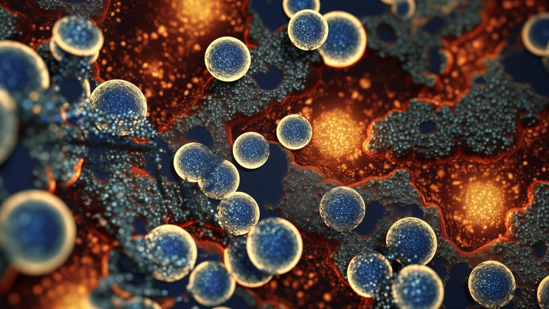
Cryo-electron microscopy (cryo-EM) is a powerful tool used to visualize the tiny structures of biological molecules in their natural state. This technique has revolutionized structural biology by allowing scientists to see proteins, viruses, and other complex molecules at near-atomic resolution without the need for crystallization. Imagine peering into the microscopic world and observing the intricate dance of molecules as they perform their vital functions. Cryo-EM achieves this by rapidly freezing samples, preserving their native structures, and then using electron beams to capture detailed images. This method has been pivotal in understanding diseases, developing new drugs, and advancing biotechnology. With its ability to reveal the unseen, cryo-EM continues to push the boundaries of what we know about the molecular world. Whether you're a budding scientist or just curious about how life works at the smallest scale, cryo-EM offers a fascinating glimpse into the building blocks of life.
Key Takeaways:
- Cryo-electron microscopy (cryo-EM) allows scientists to see tiny biological molecules in incredible detail without needing to crystallize them. It's like taking super clear pictures of things too small to see with a regular microscope!
- Cryo-EM has helped us understand viruses, develop new drugs, and study diseases like Alzheimer's. It's a powerful tool, but it's also tricky and expensive to use. Scientists are working to make it even better and more accessible.
What is Cryo-Electron Microscopy?
Cryo-electron microscopy (cryo-EM) is a groundbreaking technique in structural biology. It allows scientists to visualize proteins and other biological molecules at near-atomic resolution. This method has revolutionized our understanding of molecular structures and their functions.
-
Cryo-EM involves freezing samples: Samples are rapidly frozen to preserve their natural state. This process prevents the formation of ice crystals, which can damage delicate structures.
-
No need for crystallization: Unlike X-ray crystallography, cryo-EM does not require samples to be crystallized. This makes it easier to study proteins that are difficult to crystallize.
-
Uses electron beams: Instead of light, cryo-EM uses electron beams to capture images. Electrons have much shorter wavelengths than visible light, allowing for higher resolution imaging.
-
Nobel Prize-winning technique: In 2017, the Nobel Prize in Chemistry was awarded to Jacques Dubochet, Joachim Frank, and Richard Henderson for their work in developing cryo-EM.
How Does Cryo-EM Work?
Understanding the process of cryo-EM can be fascinating. It involves several steps, each crucial for obtaining high-resolution images of biological molecules.
-
Sample preparation is key: Samples are placed on a grid and rapidly frozen in liquid ethane. This step is crucial to maintain the sample's natural structure.
-
Imaging with electron microscopes: The frozen samples are then imaged using an electron microscope. Thousands of images are taken from different angles.
-
Image processing and reconstruction: Advanced software is used to process the images and reconstruct a 3D model of the molecule. This step requires significant computational power.
-
Resolution has improved over time: Advances in technology have significantly improved the resolution of cryo-EM images. Today, scientists can achieve near-atomic resolution.
Applications of Cryo-EM
Cryo-EM has a wide range of applications in biology and medicine. It has become an essential tool for researchers studying complex molecular structures.
-
Studying viruses: Cryo-EM has been instrumental in studying viruses, including the structure of the Zika virus and SARS-CoV-2, the virus responsible for COVID-19.
-
Drug discovery: By revealing the structures of proteins involved in diseases, cryo-EM aids in the development of new drugs. It helps researchers understand how drugs interact with their targets.
-
Understanding cellular machinery: Cryo-EM allows scientists to study large molecular complexes, such as ribosomes and proteasomes, which are essential for cellular function.
-
Insights into neurodegenerative diseases: Researchers use cryo-EM to study proteins involved in neurodegenerative diseases like Alzheimer's and Parkinson's. This can lead to better understanding and treatment options.
Challenges and Limitations
Despite its many advantages, cryo-EM also has some challenges and limitations. Understanding these can help in appreciating the complexity of this technique.
-
Sample preparation can be tricky: Preparing samples for cryo-EM requires skill and precision. Any mistakes can lead to poor-quality images.
-
High cost and complexity: Cryo-EM equipment is expensive and requires specialized training to operate. This limits its accessibility to some research institutions.
-
Radiation damage: Prolonged exposure to electron beams can damage samples. Researchers must balance between obtaining high-resolution images and minimizing damage.
-
Data processing is time-consuming: The process of reconstructing 3D models from 2D images is computationally intensive and can take a long time.
Future of Cryo-EM
The future of cryo-EM looks promising, with ongoing advancements and potential new applications. Researchers continue to push the boundaries of what this technique can achieve.
-
Improving resolution further: Scientists are working on improving the resolution of cryo-EM images even further, allowing for more detailed studies of molecular structures.
-
Automation and AI: Automation and artificial intelligence are being integrated into cryo-EM workflows, making the process faster and more efficient.
-
Expanding applications: As technology advances, cryo-EM may find new applications in fields like materials science and nanotechnology.
-
Increased accessibility: Efforts are being made to reduce the cost and complexity of cryo-EM, making it more accessible to researchers worldwide.
The Final Freeze Frame
Cryo-electron microscopy has transformed how scientists see the microscopic world. By freezing samples at ultra-low temperatures, this technique captures biological structures in their natural state. This method has been a game-changer for drug discovery, helping researchers understand how proteins and other molecules work. It's not just about pretty pictures; it's about unlocking secrets that could lead to new treatments for diseases. The resolution of cryo-EM keeps getting better, allowing for more detailed images than ever before. As technology advances, cryo-EM will likely become even more accessible and powerful. This means more discoveries and breakthroughs in biochemistry and molecular biology. Whether you're a budding scientist or just curious, cryo-EM is a fascinating field with a bright future. Keep an eye on this technology—it's bound to keep making headlines in the world of science.
Frequently Asked Questions
Was this page helpful?
Our commitment to delivering trustworthy and engaging content is at the heart of what we do. Each fact on our site is contributed by real users like you, bringing a wealth of diverse insights and information. To ensure the highest standards of accuracy and reliability, our dedicated editors meticulously review each submission. This process guarantees that the facts we share are not only fascinating but also credible. Trust in our commitment to quality and authenticity as you explore and learn with us.