


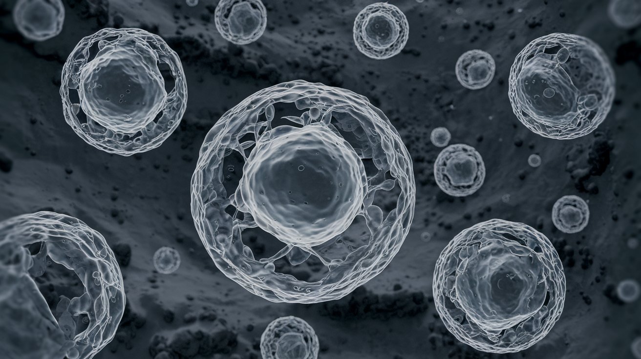
Electron microscope images reveal a world hidden from the naked eye. These powerful tools magnify objects up to two million times, allowing scientists to see details as small as atoms. Electron microscopes use beams of electrons instead of light, providing much higher resolution. This technology has transformed fields like biology, materials science, and nanotechnology. Imagine seeing the intricate structure of a virus or the detailed surface of a tiny crystal. Electron microscope images help researchers understand complex structures and processes, leading to breakthroughs in medicine, engineering, and environmental science. Ready to dive into 35 fascinating facts about these incredible images? Let's get started!
Key Takeaways:
- Explore a whole new world with electron microscope images! From butterfly wings to fruit fly eyes, these tiny details reveal a fascinating and beautiful side of science.
- Electron microscopes are like super-powered magnifying glasses, showing us the tiniest structures and textures in amazing detail. They help scientists in biology, medicine, and materials science make groundbreaking discoveries.
What is an Electron Microscope?
An electron microscope uses a beam of electrons to create an image of a specimen. This technology allows scientists to see objects at a much higher resolution than light microscopes.
- Electron microscopes can magnify objects up to 10 million times their actual size.
- The first electron microscope was developed in 1931 by Ernst Ruska and Max Knoll.
- There are two main types: Transmission Electron Microscopes (TEM) and Scanning Electron Microscopes (SEM).
How Does an Electron Microscope Work?
Understanding how these microscopes function can help appreciate the incredible images they produce.
- TEMs pass electrons through a thin specimen to form an image.
- SEMs scan a focused beam of electrons across the surface of a specimen.
- Electrons interact with atoms in the sample, producing signals that contain information about the sample's surface topography and composition.
Amazing Images Captured by Electron Microscopes
Electron microscopes reveal details invisible to the naked eye. Here are some fascinating examples.
- The intricate structure of a butterfly wing looks like a mosaic under an electron microscope.
- Pollen grains appear as spiky orbs, each unique to its plant species.
- The surface of a human hair shows scales and grooves not visible with a light microscope.
Applications in Science and Medicine
Electron microscopes play a crucial role in various scientific fields.
- In biology, they help visualize cellular structures like mitochondria and ribosomes.
- In medicine, they assist in diagnosing diseases by examining tissue samples at a microscopic level.
- Material scientists use them to study the properties of metals, polymers, and nanomaterials.
Fun Facts About Electron Microscope Images
These microscopes not only serve scientific purposes but also produce some fun and surprising images.
- The texture of a strawberry looks like a bumpy landscape.
- The surface of a CD appears as a series of tiny pits and lands.
- The eye of a fruit fly resembles a complex, multi-faceted jewel.
Challenges and Limitations
Despite their advantages, electron microscopes have some limitations.
- Samples must be placed in a vacuum, which can alter or damage them.
- Preparing specimens for electron microscopy can be time-consuming and complex.
- The equipment is expensive and requires specialized training to operate.
Innovations and Future Prospects
Advancements in electron microscopy continue to push the boundaries of what we can see.
- Cryo-electron microscopy allows scientists to observe biological specimens in their natural state.
- New techniques are improving resolution and reducing sample preparation time.
- Researchers are developing portable electron microscopes for field use.
Interesting Comparisons
Comparing electron microscope images with those from other types of microscopes highlights their unique capabilities.
- Light microscopes can only magnify objects up to 2000 times, far less than electron microscopes.
- Atomic force microscopes provide 3D surface profiles but lack the resolution of electron microscopes.
- Fluorescence microscopes can visualize specific proteins within cells, but electron microscopes reveal more structural details.
Historical Milestones
The development of electron microscopy has a rich history filled with significant achievements.
- The first commercial electron microscope was produced in 1939 by Siemens.
- In 1986, Ernst Ruska received the Nobel Prize in Physics for his work on electron optics.
- The invention of the scanning tunneling microscope in 1981 allowed scientists to visualize surfaces at the atomic level.
Unusual Uses of Electron Microscopes
Beyond traditional scientific research, electron microscopes have found some unexpected applications.
- Artists use electron microscope images to create intricate and detailed artworks.
- Forensic scientists examine tiny particles of evidence, such as gunshot residue or fibers.
- Paleontologists study the microstructures of fossils to learn about ancient life forms.
Electron Microscope Images in Popular Culture
These stunning images have also made their way into popular culture.
- Electron microscope images are often featured in science fiction movies and TV shows.
- They appear in educational materials, helping students understand complex scientific concepts.
- Some fashion designers use electron microscope images as inspiration for textile patterns.
The Future of Electron Microscopy
The field of electron microscopy continues to evolve, promising even more exciting discoveries.
- Advances in artificial intelligence are enhancing image analysis and interpretation.
- New materials and techniques are expanding the range of specimens that can be studied.
The Wonders of Electron Microscopes
Electron microscopes open up a whole new world. They let us see things way smaller than what regular microscopes can show. From tiny bugs to the smallest parts of our cells, these images are mind-blowing. Scientists use them to make big discoveries in medicine, biology, and materials science. Without electron microscopes, we’d miss out on understanding many important things about our world. They help us learn more about diseases, create new materials, and even explore space dust. So next time you see an electron microscope image, remember it’s not just a cool picture. It’s a peek into a hidden world that helps us make big strides in science and technology. Keep your curiosity alive and who knows, maybe you’ll be the next person to uncover something amazing with an electron microscope!
Frequently Asked Questions
Was this page helpful?
Our commitment to delivering trustworthy and engaging content is at the heart of what we do. Each fact on our site is contributed by real users like you, bringing a wealth of diverse insights and information. To ensure the highest standards of accuracy and reliability, our dedicated editors meticulously review each submission. This process guarantees that the facts we share are not only fascinating but also credible. Trust in our commitment to quality and authenticity as you explore and learn with us.