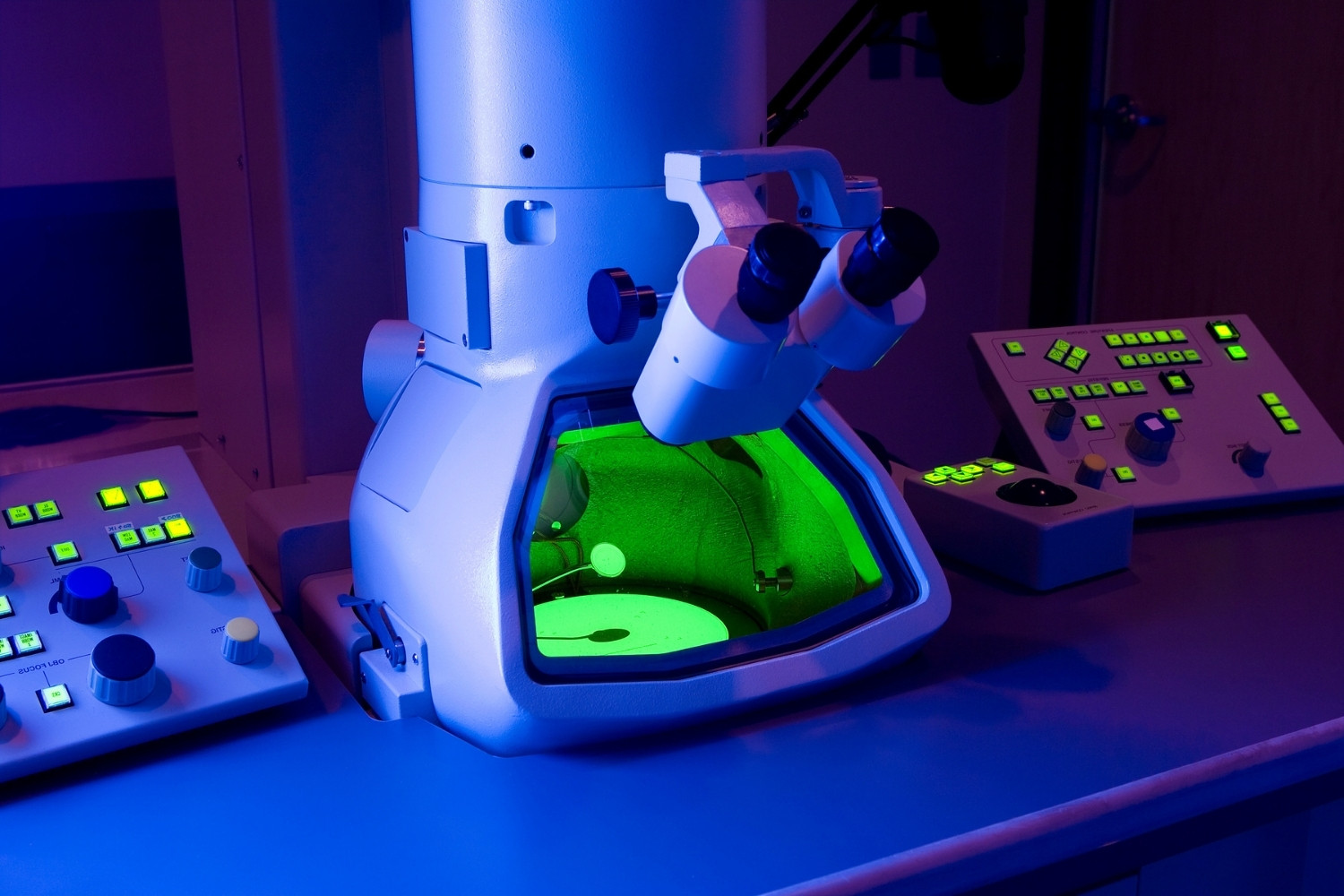
Ever wondered how scientists can see objects smaller than a cell? Electron microscopy is the answer! This powerful tool allows researchers to view tiny structures with incredible detail. Unlike regular microscopes that use light, electron microscopes use beams of electrons, which have much shorter wavelengths. This means they can reveal much smaller details. Imagine being able to see the intricate patterns on a butterfly's wing or the tiny structures inside a virus. Electron microscopy has revolutionized fields like biology, materials science, and nanotechnology. Ready to dive into some mind-blowing facts about this amazing technology? Let's get started!
What is Electron Microscopy?
Electron microscopy uses a beam of electrons to create an image of a specimen. This technique provides much higher resolution than light microscopy, allowing scientists to see tiny details.
-
Electron microscopes can magnify objects up to 10 million times. This incredible magnification allows researchers to observe the smallest structures in cells and materials.
-
There are two main types of electron microscopes: Transmission Electron Microscopes (TEM) and Scanning Electron Microscopes (SEM). TEMs transmit electrons through a specimen, while SEMs scan the surface with electrons.
-
Electron microscopes use electromagnetic lenses instead of glass lenses. These lenses focus the electron beam to create a detailed image.
-
The first electron microscope was developed in the 1930s. German engineers Ernst Ruska and Max Knoll built the first prototype in 1931.
-
Electron microscopes require a vacuum to operate. Electrons can be scattered by air molecules, so a vacuum ensures a clear path for the electron beam.
How Electron Microscopy Works
Understanding how electron microscopy works can help appreciate its capabilities. The process involves several steps to produce high-resolution images.
-
Specimens must be very thin for TEM. Thin sections allow electrons to pass through, creating a detailed image of the internal structure.
-
SEM samples are coated with a thin layer of metal. This coating enhances the electron emission from the surface, improving image quality.
-
Electron beams are generated by an electron gun. This device emits electrons that are then focused into a beam by electromagnetic lenses.
-
Detectors capture electrons to form an image. Different detectors can capture various signals, such as secondary electrons or backscattered electrons, providing different types of information.
-
Images are displayed on a computer screen. Modern electron microscopes use digital technology to display and analyze images.
Applications of Electron Microscopy
Electron microscopy has a wide range of applications in science and industry. Its ability to reveal fine details makes it invaluable in many fields.
-
Biologists use electron microscopy to study cell structures. It reveals organelles, membranes, and other cellular components in great detail.
-
Materials scientists analyze the microstructure of materials. This helps in understanding properties like strength, conductivity, and corrosion resistance.
-
Electron microscopy aids in nanotechnology research. It allows scientists to visualize and manipulate structures at the nanoscale.
-
Forensic scientists use electron microscopy for evidence analysis. It can identify tiny particles, fibers, and residues at crime scenes.
-
Geologists study mineral compositions with electron microscopy. It helps in identifying minerals and understanding geological processes.
Advantages of Electron Microscopy
Electron microscopy offers several advantages over traditional light microscopy. These benefits make it a powerful tool for scientific research.
-
Higher resolution than light microscopy. Electron microscopes can reveal structures that are too small to be seen with light microscopes.
-
Greater depth of field in SEM. This allows for detailed 3D images of surfaces.
-
Ability to analyze chemical composition. Techniques like Energy Dispersive X-ray Spectroscopy (EDS) can be used with electron microscopes to identify elements in a sample.
-
High contrast images. Electron microscopy can produce images with high contrast, making it easier to distinguish different structures.
-
Versatility in sample types. Electron microscopes can be used to study a wide range of samples, from biological tissues to metals and ceramics.
Challenges and Limitations
Despite its advantages, electron microscopy also has some challenges and limitations. Understanding these can help in effectively using this technology.
-
Sample preparation can be complex. Preparing samples for electron microscopy often requires specialized techniques and equipment.
-
Electron microscopes are expensive. The cost of purchasing and maintaining these instruments can be high.
-
Operation requires specialized training. Using an electron microscope effectively requires knowledge and experience.
-
Samples can be damaged by the electron beam. High-energy electrons can alter or destroy delicate samples.
-
Limited field of view. Electron microscopes can only image small areas at a time, which can be a limitation for studying large samples.
Innovations in Electron Microscopy
Recent innovations have expanded the capabilities of electron microscopy. These advancements continue to push the boundaries of what can be observed.
-
Cryo-electron microscopy allows imaging of biological samples in their natural state. This technique freezes samples rapidly, preserving their structure without the need for staining or fixing.
-
Environmental SEM (ESEM) enables imaging of wet or uncoated samples. ESEM can operate at higher pressures, allowing for the study of samples in more natural conditions.
-
Aberration-corrected electron microscopes offer even higher resolution. These instruments correct for lens imperfections, providing clearer images.
-
Automated electron microscopy systems increase efficiency. Automation can speed up imaging and analysis, making it easier to handle large numbers of samples.
-
Advances in detector technology improve image quality. New detectors can capture more information, providing better images and more data.
Fun Facts About Electron Microscopy
Electron microscopy isn't just about serious science. There are some fun and surprising aspects to this technology.
-
Electron microscopes can reveal the beauty of the microscopic world. Images of tiny structures can be surprisingly artistic and beautiful.
-
Some electron microscopes are used in art conservation. They help analyze and preserve historical artifacts and artworks.
-
Electron microscopy has been used to study extraterrestrial materials. Scientists have examined meteorites and moon rocks with electron microscopes.
-
The smallest things ever seen by humans were imaged with electron microscopes. This includes individual atoms and molecules.
-
Electron microscopes have contributed to Nobel Prize-winning research. Several Nobel Prizes in Chemistry and Physics have been awarded for discoveries made using electron microscopy.
Future of Electron Microscopy
The future of electron microscopy looks promising, with ongoing research and development. These advancements will continue to enhance its capabilities.
-
Development of faster and more powerful electron microscopes. Future instruments will provide even higher resolution and faster imaging.
-
Integration with other technologies. Combining electron microscopy with techniques like spectroscopy and tomography will provide more comprehensive data.
-
Improved sample preparation techniques. New methods will make it easier to prepare samples for electron microscopy.
-
Increased accessibility. Efforts are being made to make electron microscopy more affordable and accessible to a wider range of researchers.
-
Continued discoveries. As electron microscopy technology advances, it will continue to reveal new insights into the microscopic world.
The Microscopic World Awaits
Electron microscopy has revolutionized how we see the world. From revealing the intricate details of viruses to uncovering the secrets of materials science, this technology has opened up new frontiers in research. Scientists can now study structures at the atomic level, leading to breakthroughs in medicine, biology, and engineering.
Understanding these 40 facts about electron microscopy gives you a glimpse into its vast potential. Whether you're a student, a researcher, or just curious, knowing how electron microscopes work and what they can do is fascinating.
So, next time you think about the tiny world around us, remember the incredible power of electron microscopy. It’s not just about seeing small things; it’s about discovering new possibilities and pushing the boundaries of what we know. The microscopic world is full of wonders, and electron microscopy is your key to exploring it.
Was this page helpful?
Our commitment to delivering trustworthy and engaging content is at the heart of what we do. Each fact on our site is contributed by real users like you, bringing a wealth of diverse insights and information. To ensure the highest standards of accuracy and reliability, our dedicated editors meticulously review each submission. This process guarantees that the facts we share are not only fascinating but also credible. Trust in our commitment to quality and authenticity as you explore and learn with us.
