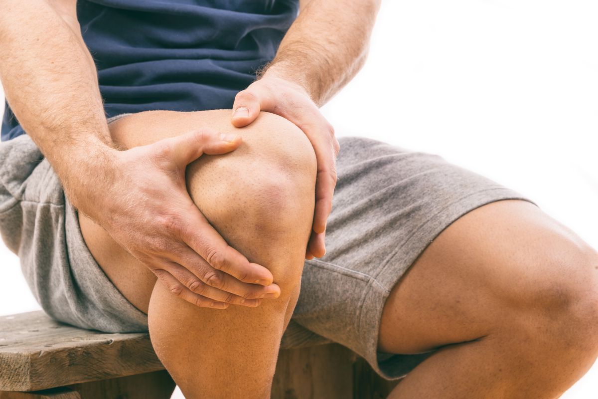
The kneecap, or patella, is a small yet vital bone at the front of the knee joint. It plays a crucial role in protecting the knee and boosting the efficiency of the quadriceps muscle. This triangular bone, embedded within the quadriceps tendon, enhances leg extension and shields the knee from physical trauma. Despite its size, the patella is the largest sesamoid bone in the body, featuring a medial and lateral facet that articulate with the femur. Understanding the anatomy, functions, and common injuries of the kneecap is essential for maintaining knee health and preventing injuries. Let's explore 45 intriguing facts about this remarkable bone.
Key Takeaways:
- The kneecap, or patella, is a small but crucial bone in the knee joint, protecting it from injury and enhancing muscle movement efficiency.
- Understanding the patella's anatomy, functions, and common injuries is essential for maintaining healthy knees and seeking timely treatment when needed.
Location and Position
The kneecap, or patella, is a small but vital bone in the knee joint. It plays a crucial role in protecting the knee and aiding movement.
- The patella is situated at the front of the knee joint, embedded within the quadriceps tendon.
- It slides along a groove on the front of the femur known as the patellofemoral groove.
Shape and Size
The patella's unique shape and size contribute to its function and efficiency.
- The patella is a triangular-shaped bone.
- Its apex is situated inferiorly and connected to the tibial tuberosity by the patellar ligament.
- The base forms the superior aspect of the bone and provides the attachment area for the quadriceps tendon.
Functions
The patella serves several important functions in the knee joint.
- Enhances the leverage that the quadriceps tendon can exert on the femur, increasing muscle movement efficiency.
- Protects the anterior aspect of the knee joint from physical trauma.
Anatomy
Understanding the patella's anatomy helps in diagnosing and treating knee issues.
- The patella is classified as a sesamoid bone due to its position within the quadriceps tendon.
- It is the largest sesamoid bone in the body.
- It has two main facets: a medial facet that articulates with the medial condyle of the femur and a lateral facet that articulates with the lateral condyle.
Bony Landmarks
Key landmarks on the patella are essential for its function and identification.
- The anterior surface is the front surface of the patella.
- The posterior surface is the back surface, which articulates with the femur.
- The medial facet is the side closer to the inside of the body.
- The lateral facet is the side closer to the outside of the body.
Attachment Points
The patella connects to other structures in the knee through various attachment points.
- It is attached to the tibia via the patellar ligament.
- It connects to the quadriceps tendon via its base.
- The quadriceps tendon links the patella to the quadriceps muscle on the front of the thigh.
Articulation
The patella's articulation with the femur is crucial for smooth knee movement.
- The patella articulates with the femur through the patellofemoral joint.
- This joint is covered with articular cartilage, providing a smooth, low-friction surface for movement.
Cartilage Coverage
The patella's cartilage plays a significant role in reducing friction and absorbing shock.
- The patella has the thickest layer of cartilage in the body.
Joint Compartments
The knee joint is divided into three compartments, each playing a role in knee function.
- The medial compartment is the joint between the femur and tibia on the inner side of the knee.
- The lateral compartment is the joint between the femur and tibia on the outer side of the knee.
- The patellofemoral compartment is the joint between the patella and its groove on the femur.
Ligaments
Several ligaments stabilize the knee joint, including those connected to the patella.
- The medial collateral ligament (MCL) connects the inner sides of the femur and tibia.
- The lateral collateral ligament (LCL) connects the outer sides of the femur and tibia.
- The anterior cruciate ligament (ACL) runs from the back of the outer condyle to the front of the tibia.
- The posterior cruciate ligament (PCL) runs from the front of the inner condyle to the back of the tibia.
Ligament Functions
These ligaments provide essential support and stability to the knee joint.
- The MCL and LCL stabilize the knee when it is straight.
- The ACL and PCL support the knee when it is bent.
Cruciate Ligament Roles
The cruciate ligaments are crucial for knee stability in various directions.
- The ACL prevents the femur from sliding backward on the tibia.
- The PCL prevents the femur from sliding forward on the tibia.
Common Injuries
The patella is susceptible to several types of injuries, affecting knee function.
- Dislocation occurs when the patella is forced out of its normal position in the femoral groove.
- Fracture can happen due to direct trauma or sudden contraction of the quadriceps muscle.
- Tendon tears can occur where the patellar tendon attaches to the bottom of the kneecap.
- Inflammation can soften and damage the cartilage underneath the patella.
Patellar Dislocation
Patellar dislocation is a common injury, especially in sports.
- It occurs when the patella is forced out of its normal position in the femoral groove.
- High-force impacts or sudden twisting of the knee can cause dislocation.
Patellar Fracture
Patellar fractures are often the result of direct trauma or muscle contraction.
- They are more common in males and in the 20-50 age range.
- Fractures can cause the patella to break into fragments, which may separate.
Symptoms of Injury
Recognizing the symptoms of a patella injury is crucial for timely treatment.
- Common symptoms include knee pain with movement, tenderness, swelling, and inability to stand or walk.
Treatment Options
Various treatments are available for patella injuries, depending on severity.
- Anti-inflammatory medications can help treat pain.
- The R.I.C.E. protocol (rest, ice, compression, elevation) reduces swelling and promotes healing.
- In severe cases, surgery may be required to correct displaced fractures or repair damaged tissues.
Rehabilitation
Rehabilitation is essential for recovering from a patella injury.
- Physical therapy helps strengthen surrounding muscles and improve joint stability.
Osteoporosis Risk
The patella, like all bones, can be affected by osteoporosis.
- Osteoporosis weakens bones, making them more susceptible to fractures.
The Kneecap's Vital Role
The kneecap, or patella, is more than just a small bone in your knee. It enhances the efficiency of the quadriceps muscle and protects the knee joint from trauma. This triangular bone, embedded within the quadriceps tendon, plays a crucial role in leg extension and movement. It's also the largest sesamoid bone in the body, with thick cartilage to reduce friction and absorb shock.
However, the patella is prone to injuries like dislocation, fracture, and tendon tears. Proper treatment and rehabilitation are essential for recovery. Understanding the patella's anatomy and functions helps in diagnosing and treating knee issues effectively.
Maintaining bone health through regular exercise and a balanced diet can prevent many patella-related problems. Despite its size, the kneecap is a key player in overall knee function and health, making it indispensable for daily activities.
Frequently Asked Questions
Was this page helpful?
Our commitment to delivering trustworthy and engaging content is at the heart of what we do. Each fact on our site is contributed by real users like you, bringing a wealth of diverse insights and information. To ensure the highest standards of accuracy and reliability, our dedicated editors meticulously review each submission. This process guarantees that the facts we share are not only fascinating but also credible. Trust in our commitment to quality and authenticity as you explore and learn with us.
