


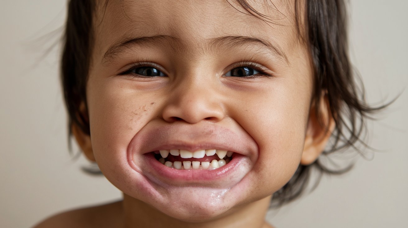
Cherubism is a rare genetic disorder that gives children a cherubic appearance due to the symmetrical enlargement of their jaw bones. This condition, first identified in 1933, affects both boys and girls equally. Caused by mutations in the SH3BP2 gene, it typically appears between ages 2 and 7. The disorder leads to painless, bilateral, multilocular radiolucent lesions in the mandible and maxilla. While most cases regress after puberty, some persist into adulthood. Diagnosis relies on clinical and radiographic findings, with genetic testing available for confirmation. Treatment focuses on managing symptoms and improving facial aesthetics, often through conservative methods.
Key Takeaways:
- Cherubism is a rare genetic disorder that affects the jaw bones, causing a cherubic appearance in children. It's caused by mutations in the SH3BP2 gene and usually improves with time, but may require surgical intervention in severe cases.
- Cherubism typically appears in early childhood, with boys slightly more affected than girls. It's diagnosed through clinical evaluation and radiographic examination, and can cause facial swelling and noticeable changes in the jaw bones.
What is Cherubism?
Cherubism is a rare genetic disorder that affects the jaw bones, giving children a cherubic appearance. This condition is marked by the symmetrical enlargement of the mandible and maxilla. Let's dive into some key facts about cherubism.
- Cherubism is a congenital childhood disease of autosomal dominant inheritance.
- It was first recognized as a separate entity in 1933 by William A. Jones.
- The name "cherubism" comes from the full round cheeks and upward cast of the eyes, resembling cherubs in baroque art.
How Common is Cherubism?
Cherubism is quite rare, with only a few hundred cases reported worldwide. It affects both genders equally and can occur in any racial or ethnic group.
- Only an estimated 300 cases of cherubism have been reported in the literature.
- It affects males and females with equal frequency.
- Cherubism has been reported in patients of all racial and ethnic backgrounds.
Genetic Basis of Cherubism
The disorder is caused by mutations in a specific gene that affects bone resorption and osteoclast activity. This leads to the characteristic changes seen in the jaw bones.
- Cherubism is caused by mutations in the SH3-binding protein 2 (SH3BP2) gene.
- These mutations lead to abnormal activation of osteoclasts.
- The abnormal activation results in the replacement of bone tissue with fibrous tissue.
Clinical Presentation of Cherubism
Cherubism typically manifests in early childhood and is characterized by painless, symmetrical lesions in the jaw bones. These lesions can cause noticeable facial changes.
- The hallmark of cherubism is the development of symmetrical multilocular radiolucent expansile lesions.
- These lesions usually appear between the ages of 2 to 7 years.
- The lesions are typically painless.
Radiographic Features of Cherubism
Radiographic examination is crucial for diagnosing cherubism. The condition has distinct radiographic features that help differentiate it from other jaw disorders.
- Cherubism manifests radiographically as bilateral, multilocular radiolucencies.
- These radiolucencies affect the posterior mandible and maxilla.
- They are often accompanied by cortical thinning and expansion of the jaw bones.
Symptoms of Cherubism
While the condition is usually painless, it can cause significant facial swelling and other symptoms that may affect the patient's appearance and function.
- Patients may experience facial swelling, particularly in the cheeks.
- The condition can cause a rounded face appearance.
- Lesions invading the orbits can cause upward tilting or displacement of the eyes.
Diagnosis of Cherubism
Diagnosing cherubism involves a combination of clinical evaluation, radiographic examination, and sometimes histopathological analysis.
- Diagnosis is primarily based on clinical and radiographic findings.
- Radiographic examination confirms the presence of bilateral, symmetrical lesions.
- Histopathological examination may be performed to rule out other conditions.
Histopathology of Cherubism
Histopathological examination provides insight into the tissue changes occurring in cherubism, helping to confirm the diagnosis.
- Histopathology reveals fibrous tissue replacing bone tissue.
- Lesions are composed of fibrous connective tissue with scattered giant cells.
- Areas of bone resorption may also be observed.
Treatment and Management of Cherubism
Managing cherubism often involves a conservative approach, with surgical intervention reserved for severe cases.
- Treatment is primarily conservative, focusing on symptom management.
- Surgical intervention may be considered for significant facial deformity or functional impairment.
- The goal of surgery is to remove fibrous tissue and restore normal jaw architecture.
Prognosis of Cherubism
Most cases of cherubism improve with time, but some may persist or worsen into adulthood.
- Most cases regress spontaneously after puberty.
- Rare instances of persistent or actively growing lesions in young adults exist.
- The condition is generally benign but can cause cosmetic and functional issues.
Age of Onset and Sex Distribution
Cherubism typically appears in early childhood and affects boys slightly more often than girls.
- Cherubism usually manifests between the ages of 2 to 7 years.
- Boys are more frequently affected than girls, with a male-to-female ratio of approximately 2:1.
- This ratio may be due to incomplete penetrance and delayed diagnosis in females.
Family History and Inheritance
Cherubism is inherited in an autosomal dominant pattern, but sporadic cases can also occur.
- Cherubism is inherited in an autosomal dominant pattern.
- A single copy of the mutated gene is sufficient to cause the condition.
- Sporadic cases have also been reported.
Radiographic Signs and Lesion Involvement
Radiographic signs are crucial for diagnosing cherubism, and lesions are usually limited to the jaw bones.
- The first radiographic signs are usually found in the region of the mandibular angle.
- Lesions may affect the development or eruption of permanent molars.
- Rare reports exist of involvement of the zygomatic arches and condyles.
Fibrous Tissue Expansion and Orbital Involvement
The fibrous tissue in cherubism can cause significant bone changes and even affect the orbits.
- Progressive lesions result in extensive bone resorption and cortical bone thinning.
- Fibrous tissue masses can expand the cortical bone, leading to facial swelling.
- When lesions invade the orbits, they can cause upward tilting or displacement of the eyes.
Genetic Testing and Clinical Variability
Genetic testing can confirm the diagnosis of cherubism, and the severity of the condition can vary widely.
- Genetic tests can identify mutations in the SH3BP2 gene.
- The severity of the disease phenotype is highly variable, even within a family.
- Patients with a mild form may develop only small symmetric lesions in the mandible.
Cherubism: A Rare Genetic Disorder
Cherubism is a rare genetic disorder that causes symmetrical enlargement of the jaw bones, giving children a cherubic appearance. It’s caused by mutations in the SH3BP2 gene, leading to abnormal bone resorption and fibrous tissue replacement. This condition usually appears between ages 2 and 7, often regressing after puberty. Diagnosis relies on clinical and radiographic findings, sometimes confirmed by genetic testing. Treatment focuses on managing symptoms and improving facial aesthetics, with surgery considered for severe cases. Though most cases resolve on their own, cherubism can cause significant cosmetic and functional issues. Understanding its clinical, radiographic, and histopathological features is crucial for accurate diagnosis and effective management. Genetic counseling can help families understand the inheritance pattern and risks. Ongoing research aims to uncover more about the disease’s pathogenesis and develop better treatments.
Frequently Asked Questions
Was this page helpful?
Our commitment to delivering trustworthy and engaging content is at the heart of what we do. Each fact on our site is contributed by real users like you, bringing a wealth of diverse insights and information. To ensure the highest standards of accuracy and reliability, our dedicated editors meticulously review each submission. This process guarantees that the facts we share are not only fascinating but also credible. Trust in our commitment to quality and authenticity as you explore and learn with us.