


What exactly is a cavernous hemangioma? These benign vascular malformations, also known as cavernous angiomas or cerebral cavernous malformations (CCMs), are clusters of abnormal, dilated blood vessels. They can appear in various parts of the body, most commonly in the brain and spinal cord. Often, they look like tiny raspberries due to their clustered capillaries. While some people with cavernous hemangiomas remain symptom-free, others may experience seizures, headaches, or even strokes. Diagnosing these lesions can be tricky, as they often go unnoticed until they cause symptoms or are found incidentally during imaging for other issues. Understanding cavernous hemangiomas is crucial for managing their potential impacts on health.
Key Takeaways:
- Cavernous hemangiomas are abnormal clusters of blood vessels in the body, often found in the brain and spinal cord. They can cause symptoms like seizures and headaches, and may require surgery for treatment.
- Genetic factors play a role in the development of cavernous hemangiomas, and ongoing research aims to improve understanding and treatment options for this condition.
What is a Cavernous Hemangioma?
Cavernous hemangiomas, also known as cavernous angiomas or cerebral cavernous malformations (CCMs), are benign vascular malformations. They can occur in various parts of the body, most commonly in the brain and spinal cord. These lesions are characterized by clusters of abnormal, hyalinized capillaries without intervening brain tissue. Let's dive into some key facts about these intriguing malformations.
-
Definition: Cavernous hemangiomas are benign vascular malformations that form during development, characterized by clusters of abnormal, hyalinized capillaries.
-
Types: These malformations are grouped into four types based on their gross and histopathologic characteristics: capillary malformations, cavernous malformations, venous malformations, and arteriovenous shunting malformations.
-
Brain Involvement: Cavernous hemangiomas in the brain are known as cerebral cavernous malformations (CCMs) and can occur at any age, but they frequently present symptomatically in the third to sixth decades of life.
Causes and Genetic Factors
Understanding the causes and genetic factors behind cavernous hemangiomas can help in diagnosing and managing the condition more effectively.
-
Etiology: These lesions can occur sporadically or in a familial pattern. Familial cases show an autosomal dominant pattern of inheritance with incomplete penetrance, linked to genetic mutations at three loci: CCM1, CCM2, and CCM3.
-
Genetic Mutations: The most common genetic mutation linked to familial cerebral cavernous malformations is a founder mutation localized to 7q (CCM1) and 7p (CCM2) in Hispanic Americans and non-Hispanic families, with CCM3 at 3q.
-
Developmental Nature: The developmental nature of these lesions is supported by the de novo development of CMs after brain biopsy and radiosurgery.
Histopathology and Symptoms
The histopathological features and symptoms of cavernous hemangiomas are crucial for understanding their impact on health.
-
Histopathology: On histological examination, cavernomas appear as abnormally dilated and hyalinized capillaries (caverns) without intervening brain tissue. Gliosis and hemosiderin deposits are seen along the margins.
-
Symptoms: Common symptoms include seizures, stroke symptoms, hemorrhages, and headaches. The type, frequency, and severity of symptoms often depend on the location of the cavernoma.
Incidence and Demographics
Knowing who is most likely to develop cavernous hemangiomas can aid in early detection and treatment.
-
Incidence: The true incidence of cavernous hemangiomas is difficult to estimate because they are frequently misdiagnosed as other venous malformations. Approximately 0.5% of the population has CCM, but only about 40% of those with malformations have symptoms.
-
Age of Presentation: Cavernous hemangiomas can appear at any age but usually occur in the third to fourth decade of life. Approximately 25% of cases occur in children.
-
Sexual Preference: There is no sexual preference for the occurrence of CCMs, but women and patients under the age of 40 are at higher risk of bleeding.
Diagnosis and Imaging
Accurate diagnosis and imaging are essential for identifying cavernous hemangiomas and planning treatment.
-
Symptomatic vs. Asymptomatic: Asymptomatic individuals usually developed the malformation sporadically, while symptomatic individuals usually have inherited the genetic mutation.
-
Diagnosis: Cavernous hemangiomas are often diagnosed incidentally during imaging for other reasons or during the evaluation of headaches, seizures, focal neurologic deficits, or symptomatic hemorrhage.
-
Imaging Challenges: These lesions can be difficult to diagnose due to their low flow state and small size. Computed tomography (CT) imaging and cerebral angiography are insensitive for detecting these lesions, earning them the term "angiographically occult".
-
MRI Detection: Magnetic resonance imaging (MRI) is more sensitive for detecting cavernous hemangiomas, particularly with post-contrast enhancement scans that reveal areas of nodular enhancement.
Risk of Bleeding and Treatment Options
Understanding the risk of bleeding and available treatment options can help manage the condition more effectively.
-
Risk of Bleeding: The risk of first symptomatic hemorrhage is extremely low (0.08% per patient-year), but once symptomatic hemorrhage has happened, there is an annual 10-fold increased risk of subsequent bleeding.
-
Annual Hemorrhage Rate: Asymptomatic familial cases are thought to have a higher annual hemorrhage rate than asymptomatic sporadic cases.
-
Location-Specific Risk: The risk of bleeding is dependent on the cavernoma's location, presence of associated developmental venous anomaly, and gender. Brainstem lesions and CCM3 familial cases with PDCD10/CCM3 mutations are associated with a greater risk of bleeding.
-
Treatment Options: Treatment plans for cavernous hemangiomas include watching and waiting, medications to manage symptoms, and surgery. Surgery is the only cure and is typically performed on lesions that have bled recently or are causing seizures.
Surgical Considerations and Techniques
Surgical intervention can be a viable option for treating cavernous hemangiomas, especially when other treatments fail.
-
Surgical Considerations: The decision to perform surgery depends on factors such as the frequency and amount of bleeding, the number of cavernomas, their location, and the presence of other endovascular abnormalities.
-
Surgical Techniques: Surgery involves making a small opening in the skull to expose the brain (craniotomy) and using microsurgery and computer image-guided surgical navigation to remove the cavernoma with minimal disruption to the brain.
-
Post-Surgical Rehabilitation: Patients who experience neurological loss due to cavernomas may require post-surgical rehabilitation to regain lost functions.
Medication Management and Incidental Findings
Medications can help manage symptoms, and incidental findings can lead to early detection.
-
Medication Management: While medications cannot directly treat cavernomas, they can help manage symptoms such as seizures and headaches.
-
Incidental Findings: Many cavernous hemangiomas are discovered incidentally during imaging for other reasons, such as headaches or seizures.
Unique Characteristics and Prevalence
Cavernous hemangiomas have unique characteristics that set them apart from other vascular malformations.
-
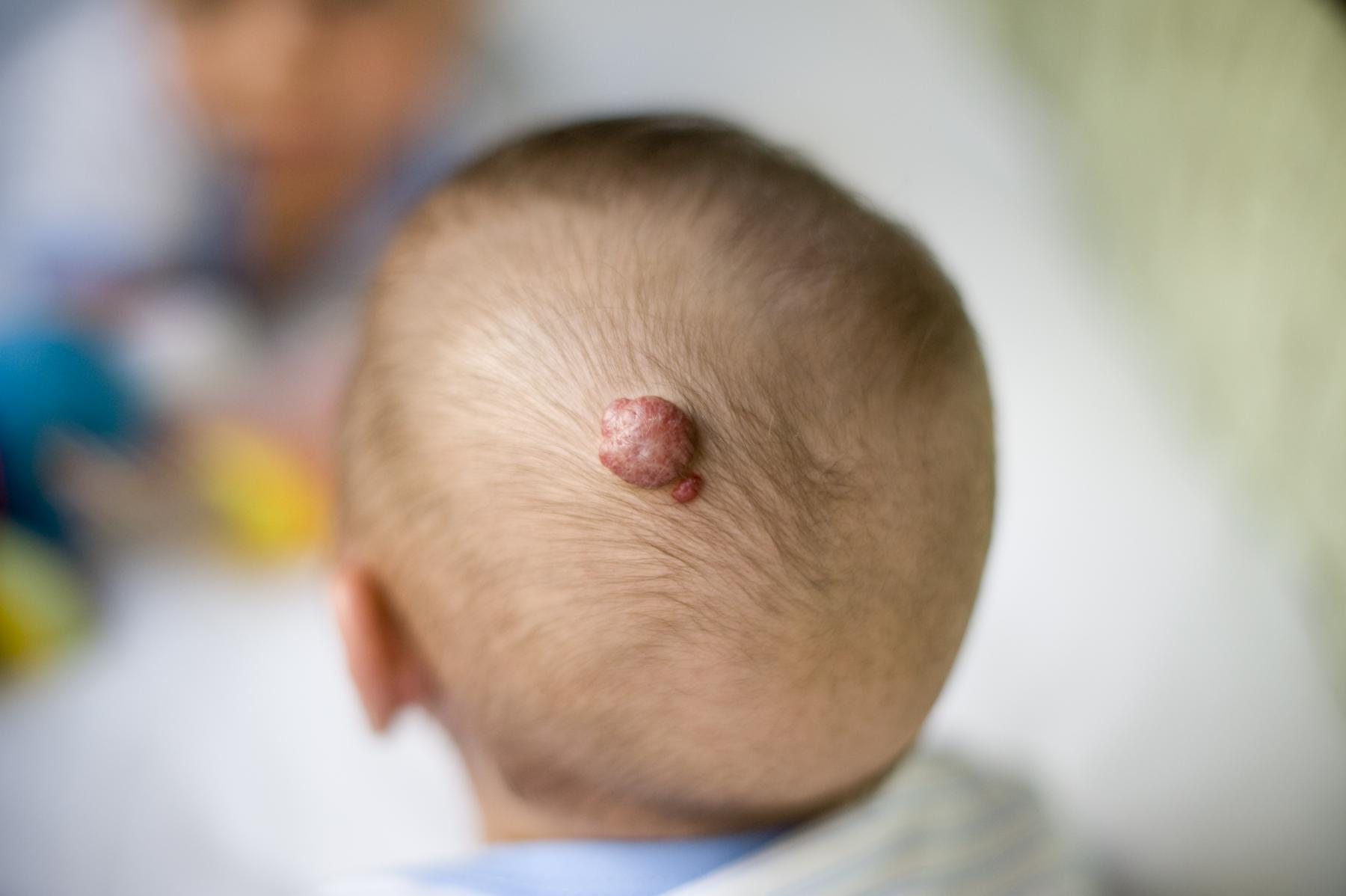
Raspberry-Shaped Appearance: Cavernomas are often described as having a raspberry-shaped appearance due to their clusters of abnormal blood vessels.
-
Leaky Walls: The walls of cavernomas are thin and leaky, which can lead to recurrent microhemorrhages and thrombosis.
-
Hemosiderin Deposits: The leakage of blood into the surrounding tissue causes hemosiderin deposits, which are seen along the margins of the lesion.
-
Gliosis: The abnormal tissue surrounding cavernomas can cause gliosis, which is an increase in glial cells in response to injury.
-
Low Flow State: Cavernomas have a low flow state, which makes them difficult to diagnose using imaging techniques like cerebral angiography.
-
Prevalence in the General Population: Approximately one in 200 people have a cavernoma, with many being present at birth and some developing later in life.
Genetic Counseling and Research
Genetic counseling and ongoing research play a crucial role in managing cavernous hemangiomas.
-
Genetic Counseling: Individuals with inherited forms of cavernomas have a 50% chance of passing the condition to each of their children. Genetic counseling can help evaluate the risk of cavernomas.
-
Research and Awareness: Organizations like the Cavernoma Alliance UK aim to raise awareness of this relatively unknown condition among the general public and the medical community.
Symptom Onset and Risk Factors
Understanding the onset of symptoms and risk factors can help in early detection and management.
-
Symptom Onset: The most common first symptom is seizure, occurring in about 50% of cases, followed by hemorrhage and focal neurological deficits.
-
Risk Factors for Bleeding: Women and patients under the age of 40 are at higher risk of bleeding from cavernous hemangiomas.
-
Annual Risk of Bleeding: The annual risk of bleeding for individuals with symptomatic cavernomas ranges from 4% to 23%, with higher risks associated with previous episodes of bleeding.
Life Expectancy and Treatment Challenges
Life expectancy and treatment challenges are important considerations for those affected by cavernous hemangiomas.
-
Life Expectancy: There is not enough data to provide a representative statistical analysis on the life expectancy of patients with cavernous hemangiomas.
-
Treatment Challenges: Cavernomas near venous malformations can make surgical treatment more difficult due to the complexity of the vascular anatomy.
-
Incidental Diagnosis: Many cavernous hemangiomas are diagnosed incidentally during imaging for other reasons, such as headaches or seizures, rather than as a primary diagnosis.
Variations in Presentation and Ongoing Research
The presentation of cavernous hemangiomas can vary widely, and ongoing research aims to improve understanding and treatment.
-
Variations in Presentation: The presentation of cavernous hemangiomas can vary widely, with some individuals experiencing no symptoms at all, while others may have severe neurological deficits.
-
Ongoing Research: Ongoing research aims to narrow down the precise mutations that cause cavernous hemangiomas and to improve diagnostic and treatment strategies for these lesions.
Final Thoughts on Cavernous Hemangiomas
Cavernous hemangiomas, or CCMs, are complex yet fascinating vascular malformations. They can appear anywhere in the body but are most concerning when found in the brain or spinal cord. Symptoms vary widely, from seizures to headaches, and sometimes they remain silent. Diagnosis often relies on MRI due to their low flow state, making other imaging techniques less effective. Treatment ranges from watchful waiting to surgical removal, depending on the lesion's location and symptoms. Genetic factors play a significant role, especially in familial cases. Understanding these malformations is crucial for effective management and improving patient outcomes. Ongoing research continues to shed light on their genetic and molecular underpinnings, paving the way for better diagnostic and treatment strategies. Stay informed and consult healthcare professionals for personalized advice.
Frequently Asked Questions
Was this page helpful?
Our commitment to delivering trustworthy and engaging content is at the heart of what we do. Each fact on our site is contributed by real users like you, bringing a wealth of diverse insights and information. To ensure the highest standards of accuracy and reliability, our dedicated editors meticulously review each submission. This process guarantees that the facts we share are not only fascinating but also credible. Trust in our commitment to quality and authenticity as you explore and learn with us.