


McConnell's sign is a fascinating medical phenomenon that can reveal a lot about heart health. Ever wondered what this sign indicates? McConnell's sign is a specific echocardiographic finding often associated with acute pulmonary embolism. It involves the presence of right ventricular dysfunction, characterized by a distinct pattern of wall motion abnormalities. This sign is named after Dr. McConnell, who first described it. Understanding McConnell's sign can be crucial for timely diagnosis and treatment of life-threatening conditions. In this blog post, we'll delve into 35 intriguing facts about McConnell's sign, shedding light on its significance, detection methods, and clinical implications. Whether you're a medical professional or just curious, these facts will enhance your knowledge about this critical diagnostic tool.
Key Takeaways:
- McConnell's Sign is a heart condition linked to lung artery blockage. It helps doctors diagnose and treat pulmonary embolism, a serious condition. Early detection can save lives!
- While McConnell's Sign is a helpful tool, it's not always present in pulmonary embolism cases. Other diagnostic methods like CT scans and blood tests can also be used.
What is McConnell's Sign?
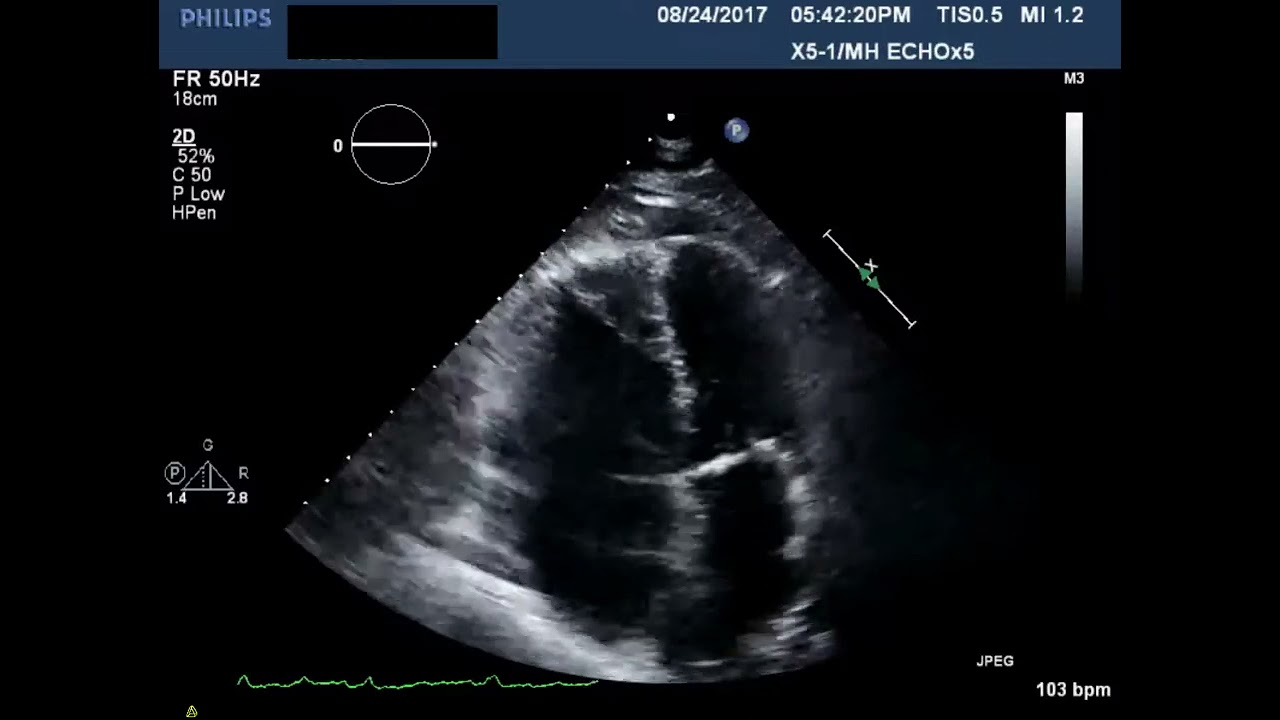
McConnell's Sign is a medical term used to describe a specific echocardiographic finding. This sign is often associated with acute pulmonary embolism, a condition where one or more arteries in the lungs become blocked by a blood clot. Understanding McConnell's Sign can help in diagnosing and treating this potentially life-threatening condition.
-
Named After Dr. McConnell: McConnell's Sign is named after Dr. Michael V. McConnell, a cardiologist who first described it in the 1990s.
-
Echocardiographic Finding: This sign is observed using an echocardiogram, a type of ultrasound that creates images of the heart.
-
Right Ventricular Dysfunction: It indicates dysfunction in the right ventricle of the heart.
-
Specific to Pulmonary Embolism: McConnell's Sign is particularly associated with acute pulmonary embolism.
-
Regional Wall Motion Abnormality: It involves a specific pattern of wall motion abnormality in the right ventricle.
How is McConnell's Sign Detected?
Detection of McConnell's Sign requires specific medical imaging techniques. These methods help doctors visualize the heart's structure and function, making it easier to identify abnormalities.
-
Transthoracic Echocardiogram: A common method for detecting McConnell's Sign is the transthoracic echocardiogram (TTE).
-
Apical Sparing: The sign is characterized by hypokinesis (reduced movement) of the mid-free wall of the right ventricle with sparing of the apex.
-
Doppler Imaging: Doppler imaging can also be used to assess blood flow and detect abnormalities.
-
Contrast Echocardiography: Sometimes, contrast agents are used to enhance the images and provide a clearer view.
-
Experienced Technicians: Accurate detection often requires experienced technicians and cardiologists.
Why is McConnell's Sign Important?
Recognizing McConnell's Sign can be crucial for timely diagnosis and treatment. Early detection can significantly improve patient outcomes.
-
Early Diagnosis: It helps in the early diagnosis of acute pulmonary embolism.
-
Guides Treatment: Identifying this sign can guide appropriate treatment strategies.
-
Prognostic Value: It has prognostic value, indicating the severity of the condition.
-
Non-Invasive: The methods used to detect McConnell's Sign are non-invasive, making it safer for patients.
-
Quick Assessment: Echocardiography allows for a quick assessment, which is vital in emergency situations.
What are the Clinical Implications?
Understanding the clinical implications of McConnell's Sign can help healthcare providers make informed decisions about patient care.
-
Risk Stratification: It helps in stratifying the risk levels of patients with suspected pulmonary embolism.
-
Monitoring: Patients with McConnell's Sign may require closer monitoring.
-
Therapeutic Decisions: It can influence therapeutic decisions, such as the need for thrombolytic therapy.
-
Hospitalization: Patients with this sign are more likely to require hospitalization.
-
Follow-Up: Regular follow-up is essential for patients diagnosed with McConnell's Sign.
How Common is McConnell's Sign?
The prevalence of McConnell's Sign can vary depending on the population and the methods used for detection.
-
Not Always Present: It is not present in all cases of pulmonary embolism.
-
Varies by Population: The prevalence can vary among different populations and settings.
-
Diagnostic Tool: It is one of several diagnostic tools used to assess pulmonary embolism.
-
Research Studies: Various research studies have investigated the prevalence and significance of McConnell's Sign.
-
Clinical Practice: Its use in clinical practice depends on the availability of echocardiography and trained personnel.
What are the Limitations?
While McConnell's Sign is a valuable diagnostic tool, it has certain limitations that should be considered.
-
False Positives: There can be false positives, where the sign is present but the patient does not have a pulmonary embolism.
-
False Negatives: Conversely, false negatives can occur, where the sign is absent despite the presence of a pulmonary embolism.
-
Operator Dependence: The accuracy of detection can depend on the skill and experience of the operator.
-
Equipment Quality: The quality of the echocardiography equipment can also affect the detection of McConnell's Sign.
-
Other Conditions: Other conditions can mimic the findings of McConnell's Sign, leading to potential misdiagnosis.
What are the Alternatives?
In cases where McConnell's Sign is not present or is inconclusive, other diagnostic methods can be used to assess pulmonary embolism.
-
CT Pulmonary Angiography: CT pulmonary angiography is a highly accurate method for diagnosing pulmonary embolism.
-
Ventilation-Perfusion Scan: A ventilation-perfusion (V/Q) scan can also be used to detect pulmonary embolism.
-
D-Dimer Test: The D-dimer blood test can help rule out pulmonary embolism in low-risk patients.
-
MRI: Magnetic resonance imaging (MRI) can be used in certain cases, although it is less common.
-
Clinical Assessment: A thorough clinical assessment, including history and physical examination, remains essential in diagnosing pulmonary embolism.
Final Thoughts on McConnell's Sign
McConnell's Sign is a fascinating and crucial indicator in the medical field. This sign, often seen in patients with acute pulmonary embolism, helps doctors quickly identify a potentially life-threatening condition. Named after Dr. Michael McConnell, it highlights the importance of echocardiography in diagnosing heart-related issues. Recognizing McConnell's Sign can lead to faster treatment and better outcomes for patients. Understanding this sign not only benefits medical professionals but also raises awareness about the seriousness of pulmonary embolism. So, next time you hear about McConnell's Sign, you'll know it's more than just a term; it's a lifesaver. Stay informed, stay healthy, and always be curious about the wonders of medical science.
Frequently Asked Questions
Was this page helpful?
Our commitment to delivering trustworthy and engaging content is at the heart of what we do. Each fact on our site is contributed by real users like you, bringing a wealth of diverse insights and information. To ensure the highest standards of accuracy and reliability, our dedicated editors meticulously review each submission. This process guarantees that the facts we share are not only fascinating but also credible. Trust in our commitment to quality and authenticity as you explore and learn with us.