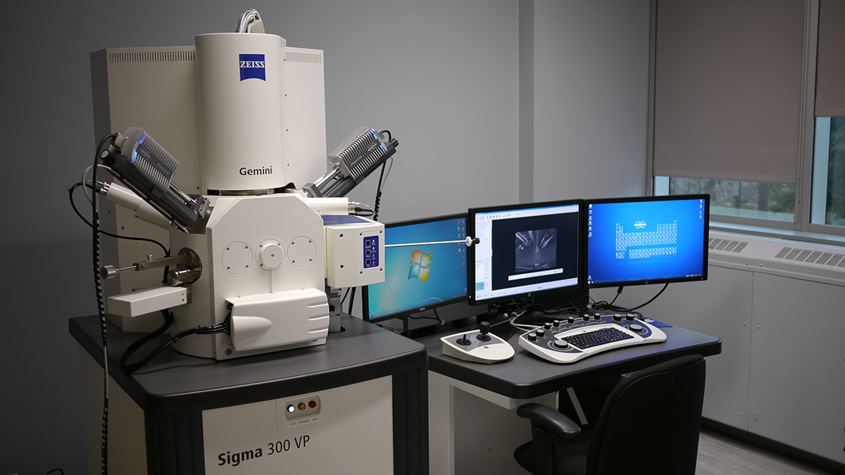
Scanning electron microscopes (SEMs) are incredible tools that let us see the world in astonishing detail. But what exactly makes them so special? SEMs use a focused beam of electrons to create detailed images of tiny objects, revealing structures invisible to the naked eye. Unlike regular microscopes, which use light, SEMs provide much higher magnification and resolution. This makes them essential in fields like biology, materials science, and nanotechnology. Ever wondered how scientists can see the surface of a tiny insect or the intricate details of a microchip? SEMs make it possible. Ready to learn more? Let's dive into 37 fascinating facts about these amazing machines!
What is a Scanning Electron Microscope (SEM)?
A Scanning Electron Microscope (SEM) is a powerful tool used to capture highly detailed images of surfaces at a microscopic level. It uses a focused beam of electrons to scan the specimen, producing images with exceptional resolution and depth of field.
-
SEM uses electrons instead of light: Unlike traditional microscopes that use light to illuminate the specimen, SEMs use a beam of electrons. This allows for much higher magnification and resolution.
-
Invented in the 1930s: The first SEM was developed by Max Knoll in 1935. However, it wasn't until the 1960s that SEMs became commercially available.
-
Magnification up to 2 million times: SEMs can magnify objects up to 2 million times their actual size, revealing details that are impossible to see with the naked eye.
How Does an SEM Work?
Understanding the working mechanism of an SEM can help appreciate its capabilities. The process involves several steps, from electron generation to image formation.
-
Electron gun generates electrons: The SEM starts with an electron gun that produces a stream of electrons. These electrons are then focused into a fine beam.
-
Electromagnetic lenses focus the beam: Electromagnetic lenses are used to focus the electron beam onto the specimen. These lenses can adjust the beam's focus and size.
-
Specimen interaction: When the electron beam hits the specimen, it interacts with the atoms on the surface, causing the emission of secondary electrons.
-
Detector captures emitted electrons: Detectors in the SEM capture the emitted secondary electrons and convert them into a signal that forms the image.
Applications of SEM
SEMs are used in various fields, from materials science to biology. Their ability to provide detailed images makes them invaluable in research and industry.
-
Materials science: SEMs are used to study the microstructure of materials, helping scientists understand properties like strength and durability.
-
Biology: In biology, SEMs can be used to examine the surface structure of cells and tissues, providing insights into their function and pathology.
-
Forensics: Forensic scientists use SEMs to analyze evidence such as gunshot residue, fibers, and hair, aiding in criminal investigations.
-
Nanotechnology: SEMs are crucial in nanotechnology for studying and manipulating materials at the nanoscale.
Advantages of Using SEM
SEMs offer several advantages over other types of microscopes, making them a preferred choice for many applications.
-
High resolution: SEMs provide images with much higher resolution compared to light microscopes, allowing for detailed observation of small structures.
-
Depth of field: The depth of field in SEM images is much greater, providing a three-dimensional appearance to the images.
-
Versatility: SEMs can be used to study a wide range of materials, from metals to biological specimens.
-
Elemental analysis: Some SEMs are equipped with energy-dispersive X-ray spectroscopy (EDS), allowing for elemental analysis of the specimen.
Challenges and Limitations of SEM
Despite their advantages, SEMs also have some limitations and challenges that users need to be aware of.
-
Sample preparation: Preparing samples for SEM analysis can be time-consuming and may require special techniques to avoid damage.
-
Vacuum environment: SEMs operate in a vacuum, which can be a limitation for studying certain types of specimens, especially those that are wet or volatile.
-
Cost: SEMs are expensive to purchase and maintain, making them less accessible for smaller laboratories or institutions.
-
Radiation damage: The electron beam can cause damage to sensitive specimens, particularly biological samples.
Interesting Facts About SEM
Here are some intriguing facts that highlight the unique capabilities and history of SEMs.
-
First commercial SEM: The first commercially available SEM was introduced by Cambridge Instrument Company in 1965.
-
SEM images are black and white: The images produced by SEMs are typically black and white, but they can be colorized using software for better visualization.
-
Cryo-SEM: Cryo-SEM is a technique where specimens are frozen to preserve their natural state during imaging.
-
Environmental SEM (ESEM): ESEM allows for the examination of specimens in a low-vacuum or wet environment, expanding the range of samples that can be studied.
-
SEM in art conservation: SEMs are used in art conservation to analyze the composition and condition of historical artifacts and artworks.
-
SEM in geology: Geologists use SEMs to study the composition and structure of minerals and rocks, helping to understand geological processes.
-
SEM in semiconductor industry: The semiconductor industry relies on SEMs for inspecting and analyzing microchips and other electronic components.
-
SEM in archaeology: Archaeologists use SEMs to examine ancient artifacts, providing insights into past civilizations and their technologies.
-
SEM in food science: Food scientists use SEMs to study the microstructure of food products, helping to improve texture and quality.
-
SEM in pharmaceuticals: In the pharmaceutical industry, SEMs are used to analyze the surface structure of drugs and other medical products.
-
SEM in environmental science: Environmental scientists use SEMs to study pollutants and their effects on the environment.
-
SEM in textiles: The textile industry uses SEMs to analyze fibers and fabrics, helping to improve material properties and performance.
-
SEM in dentistry: Dentists and researchers use SEMs to study the structure of teeth and dental materials.
-
SEM in metallurgy: Metallurgists use SEMs to examine the microstructure of metals and alloys, aiding in the development of stronger materials.
-
SEM in botany: Botanists use SEMs to study the surface structure of plants, including leaves, stems, and seeds.
-
SEM in zoology: Zoologists use SEMs to examine the surface structure of animals, including insects and other small creatures.
-
SEM in microbiology: Microbiologists use SEMs to study the surface structure of bacteria, viruses, and other microorganisms.
-
SEM in aerospace: The aerospace industry uses SEMs to analyze materials and components used in aircraft and spacecraft, ensuring their reliability and safety.
The Fascinating World of SEM
Scanning electron microscopes (SEMs) have revolutionized how we see the microscopic world. These powerful tools provide detailed images of surfaces, revealing structures invisible to the naked eye. SEMs are used in various fields like materials science, biology, and forensics, making them indispensable for research and industry.
Understanding how SEMs work helps us appreciate their impact. They use electrons instead of light to create images, allowing for much higher resolution. This technology has led to breakthroughs in nanotechnology, medicine, and environmental science.
SEMs also have practical applications, from quality control in manufacturing to analyzing crime scene evidence. Their versatility and precision make them a cornerstone of modern science and technology.
In short, SEMs open up a world of possibilities, helping us explore and understand the tiny details that make up our universe. Their importance can't be overstated.
Was this page helpful?
Our commitment to delivering trustworthy and engaging content is at the heart of what we do. Each fact on our site is contributed by real users like you, bringing a wealth of diverse insights and information. To ensure the highest standards of accuracy and reliability, our dedicated editors meticulously review each submission. This process guarantees that the facts we share are not only fascinating but also credible. Trust in our commitment to quality and authenticity as you explore and learn with us.
