


Photoacoustic imaging is a cutting-edge technology blending light and sound to create detailed images of tissues. This technique has revolutionized medical imaging, offering non-invasive ways to see inside the body. But what makes it so special? Photoacoustic imaging uses laser pulses to generate sound waves, which are then captured to form images. This method provides high-resolution images and can detect changes at the molecular level. From cancer detection to brain imaging, its applications are vast. Curious about how it works and its benefits? Here are 28 fascinating facts about photoacoustic imaging that will enlighten you on this remarkable technology.
What is Photoacoustic Imaging?
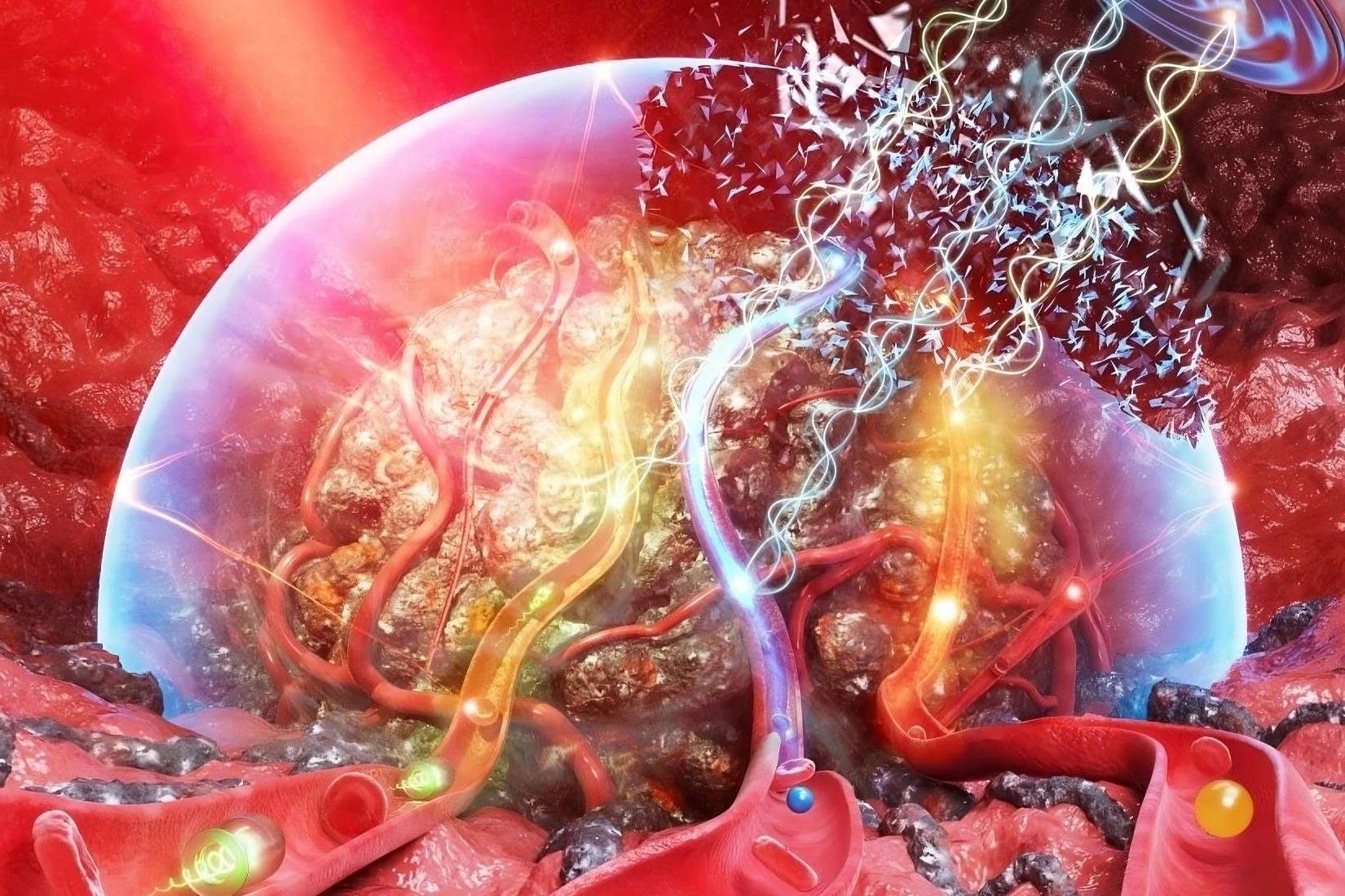
Photoacoustic imaging (PAI) is a hybrid imaging technique that combines laser-induced ultrasound with optical imaging. It provides high-resolution images of tissues, making it a valuable tool in medical diagnostics and research.
-
PAI uses laser pulses to generate ultrasound waves. When tissues absorb laser light, they heat up and expand, creating ultrasound waves that can be detected and used to form images.
-
It offers high spatial resolution. Unlike traditional ultrasound, PAI provides detailed images at a microscopic level, allowing for precise visualization of tissue structures.
-
PAI is non-invasive. This technique does not require any surgical procedures, making it safer and more comfortable for patients.
Applications in Medicine
PAI has numerous applications in the medical field, from cancer detection to monitoring blood oxygen levels. Its versatility makes it a powerful tool for various diagnostic purposes.
-
PAI can detect cancerous tumors. By highlighting differences in tissue composition, PAI helps identify malignant growths at an early stage.
-
It monitors blood oxygenation. PAI can measure the oxygen levels in blood vessels, which is crucial for assessing tissue health and detecting conditions like hypoxia.
-
PAI aids in brain imaging. Researchers use PAI to study brain function and structure, providing insights into neurological disorders.
-
It assists in cardiovascular research. PAI helps visualize blood vessels and assess their health, aiding in the diagnosis of cardiovascular diseases.
Advantages Over Other Imaging Techniques
PAI stands out due to its unique combination of optical and acoustic imaging, offering several advantages over traditional methods.
-
PAI provides deeper tissue penetration. Unlike optical imaging alone, PAI can penetrate deeper into tissues, offering more comprehensive images.
-
It offers better contrast. PAI can differentiate between various tissue types based on their optical absorption properties, providing clearer images.
-
PAI reduces the need for contrast agents. Traditional imaging techniques often require contrast agents to enhance images, but PAI can achieve high contrast without them.
Technological Advancements
Recent advancements have significantly improved the capabilities and applications of PAI, making it more accessible and effective.
-
Portable PAI devices are now available. These devices allow for bedside imaging, making it easier to monitor patients in real-time.
-
Artificial intelligence enhances PAI. AI algorithms improve image reconstruction and analysis, providing more accurate and detailed results.
-
Multi-wavelength PAI offers better imaging. Using multiple wavelengths of laser light, PAI can provide more comprehensive information about tissue composition.
Challenges and Limitations
Despite its many advantages, PAI also faces certain challenges and limitations that researchers are working to overcome.
-
PAI requires expensive equipment. The high cost of lasers and ultrasound detectors can limit its accessibility in some medical settings.
-
It has limited penetration depth. While PAI penetrates deeper than optical imaging, it still has limitations compared to traditional ultrasound.
-
PAI can be affected by tissue heterogeneity. Variations in tissue composition can impact the quality of PAI images, making it challenging to interpret results accurately.
Future Prospects
The future of PAI looks promising, with ongoing research and development aimed at expanding its applications and improving its effectiveness.
-
Researchers are exploring new contrast agents. These agents could enhance PAI images, making it easier to distinguish between different tissue types.
-
PAI may be integrated with other imaging techniques. Combining PAI with MRI or CT scans could provide even more detailed and comprehensive images.
-
Wearable PAI devices are in development. These devices could allow for continuous monitoring of patients, providing real-time data on their health.
-
PAI could revolutionize cancer treatment. By providing detailed images of tumors, PAI could help guide more precise and effective treatments.
Interesting Facts About PAI
Here are some intriguing facts about PAI that highlight its unique capabilities and potential.
-
PAI can image single cells. The high resolution of PAI allows researchers to visualize individual cells, providing insights into cellular processes.
-
It can detect melanin in the skin. PAI can differentiate between melanin and other skin components, making it useful for studying skin conditions.
-
PAI is used in plant research. Researchers use PAI to study plant tissues and monitor their health, aiding in agricultural research.
-
It can monitor drug delivery. PAI helps track the distribution of drugs within the body, ensuring they reach their intended targets.
-
PAI can image blood flow. By visualizing blood vessels, PAI provides information on blood flow dynamics, which is crucial for understanding various medical conditions.
-
It aids in wound healing studies. PAI helps researchers study the healing process of wounds, providing insights into how different treatments affect recovery.
-
PAI can detect microcalcifications. These tiny calcium deposits in tissues can be early indicators of diseases like breast cancer.
-
It is used in preclinical studies. PAI is a valuable tool in animal studies, helping researchers understand disease mechanisms and test new treatments.
The Future of Photoacoustic Imaging
Photoacoustic imaging is changing the way we see the world of medical diagnostics. This technology combines light and sound to create detailed images of tissues and organs, offering a non-invasive way to detect diseases early. Researchers are continuously improving its accuracy and applications, making it a promising tool for the future.
From detecting cancer to monitoring brain activity, the potential uses are vast. As technology advances, we can expect even more breakthroughs in this field. The ability to see inside the body without surgery opens up new possibilities for patient care and treatment.
In short, photoacoustic imaging is a game-changer in medical science. Its ongoing development promises to bring about significant improvements in how we diagnose and treat various health conditions. Keep an eye on this exciting field; it's only going to get better.
Was this page helpful?
Our commitment to delivering trustworthy and engaging content is at the heart of what we do. Each fact on our site is contributed by real users like you, bringing a wealth of diverse insights and information. To ensure the highest standards of accuracy and reliability, our dedicated editors meticulously review each submission. This process guarantees that the facts we share are not only fascinating but also credible. Trust in our commitment to quality and authenticity as you explore and learn with us.