
What is Coats Disease? Coats Disease is a rare eye condition that primarily affects young males, leading to vision problems. It involves abnormal blood vessels in the retina, causing fluid leakage and potential retinal detachment. This disorder is usually unilateral, meaning it affects only one eye. Symptoms can include blurred vision, eye strain, and in severe cases, significant vision loss. Early diagnosis and treatment are crucial to manage the condition and prevent complications. Treatment options range from laser photocoagulation to surgery, depending on the disease's stage. Understanding Coats Disease can help in recognizing symptoms early and seeking appropriate medical care.
Key Takeaways:
- Coats disease is a rare eye condition that affects the retina, primarily in young males. Early diagnosis and treatment are crucial for managing this challenging condition effectively.
- The primary features of Coats disease include retinal telangiectasia, exudative retinopathy, and potential vision loss. Treatment options range from laser therapy to surgical intervention, with varying outcomes based on the disease stage.
What is Coats Disease?
Coats disease is a rare eye condition that primarily affects the retina. It often leads to vision problems and can be challenging to diagnose and treat. Here are some key facts to help you understand this condition better.
-
Definition: Coats disease is an idiopathic retinal vascular disorder characterized by retinal telangiectasia and intraretinal or subretinal exudation without appreciable vitreoretinal traction.
-
Etiology: The exact cause of Coats disease is unknown. It is considered idiopathic, meaning it arises spontaneously without a clear reason.
-
Incidence: This condition is rare, with an estimated incidence of 0.09 per 100,000 people in the United Kingdom.
Who is Affected by Coats Disease?
Understanding who is most likely to be affected by Coats disease can help in early diagnosis and treatment.
-
Gender Prevalence: Coats disease predominantly affects males, with a male-to-female ratio of at least 3:1.
-
Age of Onset: It typically affects young males, with most cases diagnosed in the first or second decade of life. However, it can also occur in adults, particularly middle-aged males.
Symptoms and Clinical Presentation
Recognizing the symptoms early can lead to better management of Coats disease.
-
Clinical Presentation: The condition often presents with unilateral vision loss, strabismus, or leukocoria (a white pupillary reflex).
-
Retinal Changes: The primary retinal changes include telangiectasia, which is the dilation and twisting of retinal capillaries. This leads to increased permeability and leakage of fluid and blood into the retina.
-
Exudation: Leakage from the abnormal vessels results in intraretinal and/or subretinal exudation. These exudates can cause retinal detachment and vision loss.
Stages of Coats Disease
Coats disease progresses through several stages, each with its own set of challenges and symptoms.
-
Stage 1: Telangiectasia with minimal changes to the retina and vision.
-
Stage 2: Telangiectasia and exudates, leading to increased changes in the retina and potential vision loss.
-
Stage 3: Large areas of retinal swelling with possible retinal detachment.
-
Stage 4: Complete retinal detachment and glaucoma.
-
Stage 5: The eye has no sight, and there are no treatment options to improve vision.
Diagnosis and Treatment
Early diagnosis and appropriate treatment are crucial for managing Coats disease effectively.
-
Symptoms: Early symptoms may include blurred vision, eye strain, and in severe cases, significant vision loss. Leukocoria or strabismus can also be presenting signs, especially in children.
-
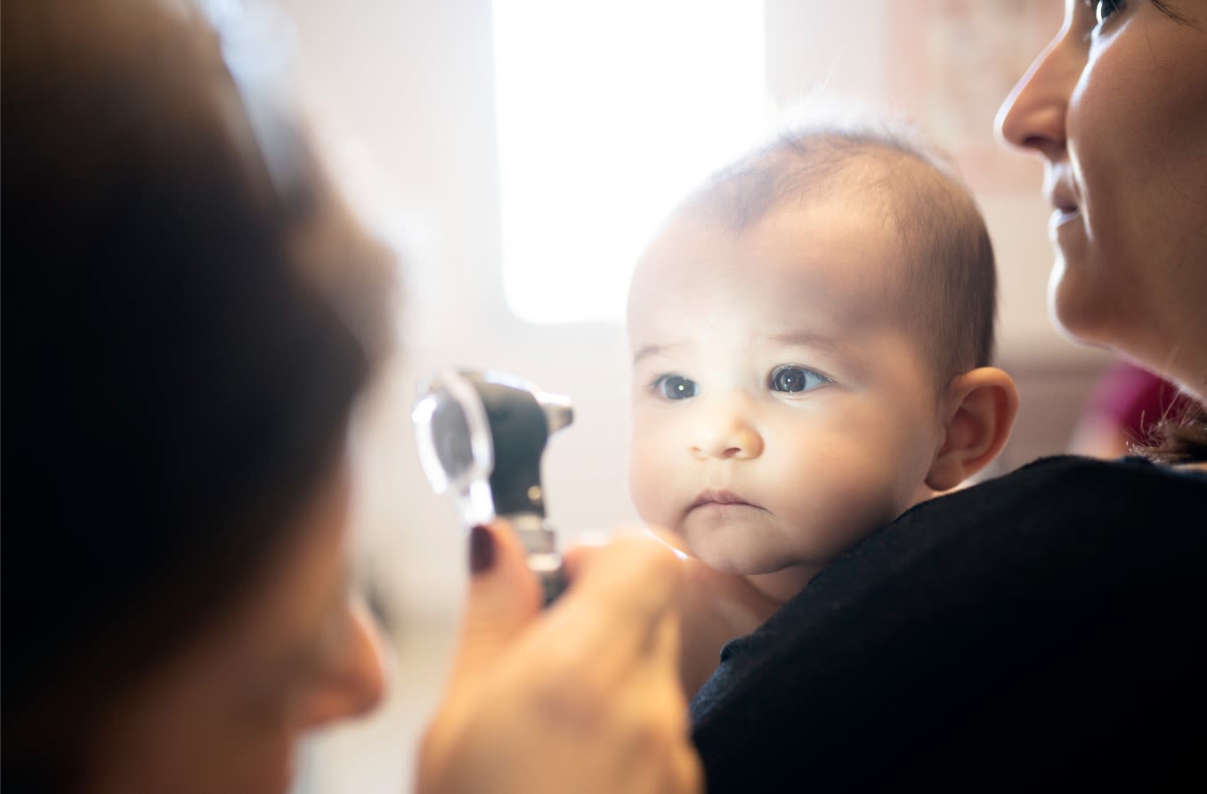
Diagnosis: Diagnosis is typically made through a comprehensive eye examination, including funduscopy and fluorescein angiography. Ancillary investigations such as optical coherence tomography (OCT) and ultrawidefield imaging can also be helpful.
-
Treatment: Management of Coats disease varies based on the stage of the disease. Initial treatments include laser photocoagulation, cryotherapy, and surgery for retinal detachment. Anti-vascular endothelial growth factor (anti-VEGF) agents have become an important adjunctive treatment.
Treatment Options
Different treatment options are available depending on the stage and severity of the disease.
-
Laser Photocoagulation: This is a common treatment for Coats disease, particularly in early stages. It involves using laser to close the abnormal vessels and reduce exudation.
-
Cryotherapy: Cryotherapy involves freezing the abnormal vessels to reduce their permeability and prevent further leakage.
-
Surgery: Surgical intervention may be necessary for advanced cases, including retinal detachment repair and vitreoretinal surgery.
-
Anti-VEGF Agents: These agents, such as bevacizumab, have been shown to be effective in reducing exudation and improving visual outcomes in Coats disease.
Visual Outcomes and Complications
The visual prognosis and potential complications can vary widely among patients.
-
Visual Outcomes: The visual prognosis varies based on the stage of the disease. Early intervention can improve outcomes, but advanced cases often result in significant vision loss.
-
Complications: Complications of Coats disease include retinal detachment, glaucoma, and neovascular glaucoma. In severe cases, enucleation may be necessary to prevent further complications.
Genetic and Histopathological Features
While the exact cause remains unknown, some genetic and histopathological features have been identified.
-
Genetic Implications: There is no known genetic basis for Coats disease. However, chromosomal instability in chromosomes 3 and 13 has been reported, and other genes have been implicated in its pathogenesis.
-
Histopathological Features: George Coats first described the histopathological features of enucleated eyes with massive exudation in 1908. He divided the morphology into three groups: massive subretinal exudate alone, massive subretinal exudate with intra and subretinal hemorrhage and retinal vascular dilatations, and subretinal exudate with retinal arteriovenous malformations.
Classification and Variants
Coats disease can be classified into different groups based on its clinical features and severity.
-
Classification: The condition is classified based on its clinical features and the extent of retinal involvement. It is often divided into three groups based on the severity of exudation and retinal changes.
-
Leber’s Miliary Aneurysm: This is considered a milder variant of Coats disease, characterized by multiple retinal aneurysms without massive subretinal exudation or hemorrhage.
-
Type 1 Idiopathic Macular Telangiectasia: This is now considered the same disease as Coats disease, characterized by telangiectatic vessels in the macula.
Retinal and Exudative Features
The primary features of Coats disease involve changes in the retinal vessels and exudation.
-
Retinal Telangiectasia: The primary pathological feature of Coats disease is retinal telangiectasia, which involves the abnormal dilation and twisting of retinal capillaries.
-
Exudative Retinopathy: The condition leads to exudative retinopathy, characterized by the leakage of fluid and blood into the retina, resulting in retinal detachment and vision loss.
-
Vitreoretinal Traction: Unlike other retinal disorders, Coats disease does not involve significant vitreoretinal traction, which makes it distinct from other conditions like retinal detachment due to trauma or disease.
Population-Based Studies and Clinical Features
Studies have provided valuable insights into the incidence and clinical features of Coats disease.
-
Population-Based Studies: A prospective population-based study in the United Kingdom identified 55 cases of Coats disease, providing insights into the incidence and clinical features of the condition in a general population.
-
Clinical Features at Presentation: The study found that most cases presented with unilateral involvement, with 85% of cases being male. The mean age at presentation was 12 years, and the majority of eyes had poor visual acuity at diagnosis.
-
Disease Severity: The study noted that more severe forms of Coats disease were more common in younger patients, indicating a poorer visual prognosis in this age group.
Treatment Preferences and Outcomes
Over the years, treatment preferences and outcomes for Coats disease have evolved.
-
Treatment Preferences: The study highlighted the importance of early diagnosis and treatment. Over the years, there has been a shift from enucleation to more conservative treatments like laser photocoagulation, cryotherapy, and anti-VEGF therapy.
-
Anatomic and Visual Outcomes: The study provided detailed information on anatomic and visual outcomes following different treatment modalities, which is crucial for clinicians planning treatment for patients with Coats disease.
Diagnostic Challenges and Historical Background
Despite its well-described clinical features, Coats disease remains a diagnostic challenge.
-
Diagnostic Challenges: Despite its well-described clinical features, Coats disease remains a diagnostic challenge due to its varied presentation and resemblance to other vascular and exudative retinopathies.
-
Historical Background: George Coats first described the condition in 1908 as unilateral retinal vascular abnormality with exudation occurring in young males. Later, Leber reported another entity characterized by multiple retinal aneurysms associated with retinal degeneration.
-
Reese’s Refinement: In 1956, Reese refined the definition and description of Coats disease, distinguishing it from other retinal conditions like Leber’s miliary aneurysms.
Coats’ Disease Spectrum and Imaging
Coats disease is part of a spectrum of retinal telangiectasias, and modern imaging techniques have improved diagnosis and management.
-
Coats’ Disease Spectrum: Coats disease is part of a spectrum of retinal telangiectasias, which includes other conditions like branch retinal vein occlusion, retinitis pigmentosa, ocular toxoplasmosis, and morning glory anomaly.
-
Ultra-Widefield Imaging: Ultra-widefield imaging and optical coherence tomography angiography have refined the diagnosis and management of Coats disease by providing detailed images of retinal vessels and exudates.
Understanding Coats Disease
Coats disease is a rare eye condition that primarily affects young males, leading to vision loss if not treated early. Characterized by abnormal retinal blood vessels and fluid leakage, it progresses through several stages, from mild telangiectasia to severe retinal detachment. Early symptoms include blurred vision and leukocoria, often prompting a visit to the eye doctor. Diagnosis involves comprehensive eye exams, including funduscopy and fluorescein angiography. Treatment options like laser photocoagulation, cryotherapy, and anti-VEGF agents can manage the condition, especially when caught early. Advanced cases might require surgery. Despite its challenges, understanding Coats disease and its treatments can significantly improve outcomes. Early intervention remains key to preserving vision and preventing complications.
Frequently Asked Questions
Was this page helpful?
Our commitment to delivering trustworthy and engaging content is at the heart of what we do. Each fact on our site is contributed by real users like you, bringing a wealth of diverse insights and information. To ensure the highest standards of accuracy and reliability, our dedicated editors meticulously review each submission. This process guarantees that the facts we share are not only fascinating but also credible. Trust in our commitment to quality and authenticity as you explore and learn with us.