

What is a chemodectoma? Chemodectomas, also known as paragangliomas, are rare, slow-growing tumors that arise from chemoreceptor cells in various parts of the body. These tumors are generally benign but can be locally invasive and, in rare cases, malignant. They typically occur around the carotid bifurcation, aortic root, main pulmonary arteries, and jugular veins. Symptoms often remain unnoticed for a long time, but when they do appear, they can include pain, discomfort, and signs related to compression of nearby structures. Diagnosing and treating chemodectomas can be challenging due to their location near critical structures like the carotid artery and cranial nerves. Treatment options include surgical excision, radiosurgery, and observation.
Key Takeaways:
- Chemodectomas, also known as paragangliomas, are rare non-functional tumors that can occur in various parts of the body. They are slow-growing and can be challenging to treat due to their location near critical structures.
- Diagnosis and treatment of chemodectomas require careful consideration of the tumor's size, location, and the patient's overall health. Surgical excision, radiosurgery, and observation are options, with ongoing research aiming to improve management strategies.
What is Chemodectoma?
Chemodectomas, also known as paragangliomas, are rare tumors that arise from chemoreceptor cells. These cells are found in various parts of the body, including the carotid bifurcation, aortic root, main pulmonary arteries, and jugular veins. Let's dive into some key facts about these intriguing tumors.
-
Definition: Chemodectomas are space-occupying masses that displace and compress adjacent structures. They are non-functional and usually benign.
-
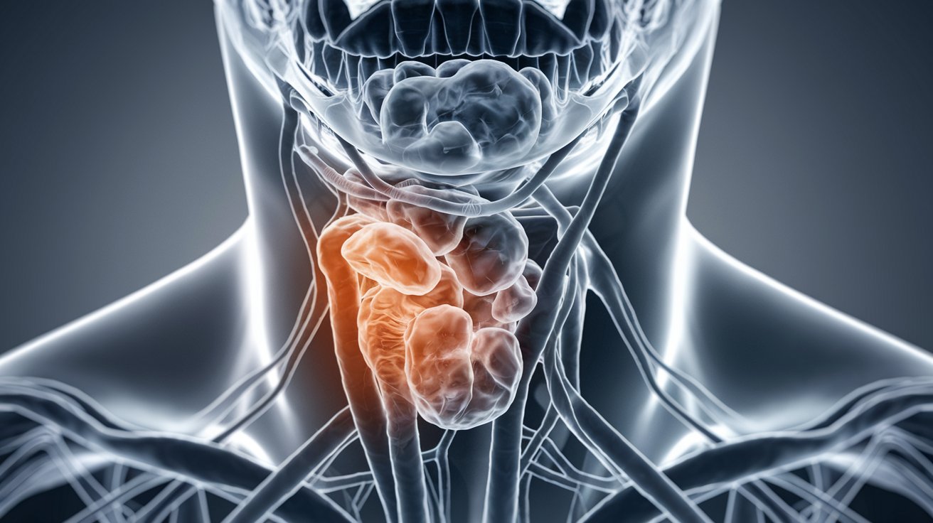
Location: These tumors typically occur around the carotid bifurcation, aortic root, main pulmonary arteries, and jugular veins.
-
Types: Different types of chemodectomas include carotid body tumors, glomus jugulare tumors, and glomus vagale tumors.
-
Incidence: Chemodectomas are relatively rare, with most cases being benign and unilateral.
Symptoms and Diagnosis
Understanding the symptoms and how these tumors are diagnosed is crucial for early detection and treatment.
-
Symptoms: Patients often remain asymptomatic for a long time, but symptoms can include pain, discomfort, and signs related to compression of nearby structures.
-
Growth Pattern: These tumors are slow-growing and can remain encapsulated, but they can also be locally invasive.
-
Histological Appearance: Histologically, chemodectomas are characterized by chief cells arranged in a "zellballen" pattern, a hallmark of paragangliomas.
-
Radiological Features: On imaging, chemodectomas appear as soft nodular, encapsulated, homogeneous masses. Various echocardiogram views help evaluate their extension and relationship with surrounding structures.
-
Diagnosis: Diagnosis is typically made through a combination of clinical examination, imaging studies (such as CT or MRI scans), and histopathological examination of biopsy samples.
Treatment Options
Treatment for chemodectomas varies based on several factors, including the tumor's size, location, and the patient's overall health.
-
Treatment Options: Options include surgical excision, radiosurgery, and observation. The choice depends on the tumor's size, location, and the patient's overall health.
-
Surgical Challenges: Surgical excision can be challenging due to the tumor's location near critical structures like the carotid artery and cranial nerves. The goal is to achieve complete tumor removal while minimizing morbidity.
-
Radiosurgery: Radiosurgery, such as CyberKnife radiotherapy, is a promising approach for managing paragangliomas. It provides high accuracy and precision, allowing for high radiation doses to the tumor while sparing healthy tissue.
-
Malignant Potential: Approximately 3-5% of chemodectomas can potentially transform into malignant tumors. The risk of malignant transformation is a significant concern in the management of these tumors.
Patient Demographics and Tumor Characteristics
Chemodectomas can affect anyone, but certain demographics are more commonly affected.
-
Patient Age: Chemodectomas can occur at any age, but they are more commonly diagnosed in middle-aged to older adults.
-
Gender Distribution: There is no significant gender predilection for chemodectomas, although some studies suggest a slight male predominance.
-
Multicentricity: While most chemodectomas are solitary, they can sometimes be multiple, particularly in cases associated with other endocrine tumors.
-
Association with Other Tumors: Chemodectomas can be associated with other endocrine tumors, such as pheochromocytomas, which are tumors of the adrenal glands.
-
Incidental Findings: Many chemodectomas are incidental findings discovered during routine echocardiograms, highlighting the importance of thorough imaging in the diagnosis of these tumors.
Complications and Prognosis
Complications can arise from both the tumor itself and its treatment. Understanding these can help manage patient expectations and outcomes.
-
Compression Symptoms: Symptoms related to compression of nearby structures, such as the pulmonary arteries or veins, can occur and may necessitate intervention.
-
Local Invasion: Chemodectomas are typically locally invasive, meaning they grow into surrounding tissues but do not metastasize to distant organs.
-
Metastasis Risk: While rare, metastasis can occur in malignant chemodectomas, complicating the management and prognosis of these cases.
-
Histological Variability: The histological appearance of chemodectomas can vary, with some tumors showing more aggressive features like increased cellularity or mitotic activity.
-
Radiation Sensitivity: The chief cells of chemodectomas are minimally affected by radiation, but the vascular structure of the tumor can be replaced by fibrous connective tissue, which is a concern in potentially malignant tumors.
-
Predictive Factors: Predictive factors like those proposed by Chapman et al. are used to assess the risk of malignant transformation in chemodectomas.
Treatment Outcomes and Future Directions
The outcomes of treatment and ongoing research are vital for improving patient care and developing new strategies.
-
Treatment Outcomes: The likelihood of tumor control is high with surgical or radiotherapeutic treatments, but there is currently no consensus on the best treatment option for all cases.
-
Individualized Treatment: Treatment must be individualized, taking into account the patient’s age, tumor site and size, multicentricity, and preexisting cranial nerve deficits.
-
Complex Cases: In selected complex cases, the combination of surgery and radiosurgery may allow complete local tumor control with minimal morbidity.
-
Cranial Nerve Involvement: Chemodectomas can involve cranial nerves, particularly those near the tumor's location, which can lead to neurological symptoms like hearing loss or facial weakness.
Specific Types of Chemodectomas
Different types of chemodectomas have unique characteristics and implications for treatment.
-
Carotid Body Tumors: These arise from the carotid body, a chemoreceptor located at the bifurcation of the carotid artery.
-
Glomus Jugulare Tumors: These arise from the jugular bulb and can extend into the middle ear and mastoid.
-
Glomus Vagale Tumors: These arise from the vagus nerve and can occur in the neck or thorax, often presenting with symptoms related to compression of nearby structures.
Historical Background and Clinical Presentation
Understanding the history and clinical presentation of chemodectomas can provide context for their diagnosis and treatment.
-
Historical Background: The carotid body was first described by von Haller in 1743, and since then, there has been significant understanding of these tumors and their management.
-
Clinical Presentation: Patients often present with a slowly growing neck mass or other symptoms related to compression of nearby structures.
-
Diagnostic Imaging: Diagnostic imaging plays a crucial role in the diagnosis of chemodectomas, including CT scans, MRI scans, and echocardiograms to evaluate the tumor's size, location, and relationship with surrounding structures.
-
Biopsy and Histopathology: Biopsy and histopathological examination are essential for confirming the diagnosis and assessing the tumor's histological features, which can guide treatment decisions.
Surgical and Radiosurgical Techniques
The techniques used to treat chemodectomas are advanced and require precision to avoid complications.
-
Surgical Techniques: Surgical techniques for chemodectoma removal involve careful dissection to avoid damaging nearby structures like the carotid artery and cranial nerves.
-
Radiosurgical Techniques: Radiosurgical techniques, such as CyberKnife radiotherapy, use precise radiation delivery to target the tumor while sparing surrounding healthy tissue.
-
Observation and Monitoring: In some cases, observation and monitoring may be recommended, especially for small, asymptomatic tumors, to avoid unnecessary interventions.
-
Complications and Morbidity: Surgical and radiosurgical interventions can have complications and morbidity, such as cranial nerve deficits or radiation-induced side effects, which must be carefully managed.
-
Research and Future Directions: Ongoing research aims to improve our understanding of chemodectomas, including their pathogenesis, molecular biology, and optimal treatment strategies, which will help in developing more effective management plans for these tumors.
Final Thoughts on Chemodectoma
Chemodectomas, also called paragangliomas, are rare, slow-growing tumors that can be tricky to diagnose and treat. They often pop up around the carotid bifurcation, aortic root, main pulmonary arteries, and jugular veins. Most of these tumors are benign, but they can be locally invasive and, in rare cases, malignant. Symptoms might not show up for a long time, making early detection tough. Treatment options include surgery, radiosurgery, and sometimes just keeping an eye on things. Each case is unique, so doctors tailor treatments to fit the patient's needs. Understanding these tumors better helps in managing them effectively. Ongoing research aims to improve our knowledge and treatment strategies, offering hope for better outcomes. Chemodectomas may be rare, but knowing the facts can make a big difference in handling them.
Frequently Asked Questions
Was this page helpful?
Our commitment to delivering trustworthy and engaging content is at the heart of what we do. Each fact on our site is contributed by real users like you, bringing a wealth of diverse insights and information. To ensure the highest standards of accuracy and reliability, our dedicated editors meticulously review each submission. This process guarantees that the facts we share are not only fascinating but also credible. Trust in our commitment to quality and authenticity as you explore and learn with us.