

Cavernous lymphangioma, also known as cavernous lymphatic malformation, is a rare but benign condition involving abnormal lymphatic vessel formation. These malformations can appear in various body parts, including the skin, subcutaneous tissues, and internal organs. What makes cavernous lymphangiomas unique? They are characterized by large, dilated lymphatic spaces. While they are more common in children and infants, adults can also develop them. Symptoms vary based on the lesion's size and location, ranging from asymptomatic to causing respiratory issues or infections. Understanding this condition's complexities is crucial for effective diagnosis and treatment.
Key Takeaways:
- Cavernous lymphangioma is a rare condition involving abnormal lymphatic vessel formation, which can appear in various body parts. Understanding its types, symptoms, and treatment options is crucial for effective management.
- Accurate diagnosis and early treatment of cavernous lymphangioma are essential for a good prognosis. Surgical excision is often the preferred treatment, but observation and sclerotherapy may also be considered based on the size and symptoms of the lesion.
What is Cavernous Lymphangioma?
Cavernous lymphangioma, also known as cavernous lymphatic malformation, is a rare, benign condition involving abnormal lymphatic vessel formation. These malformations can appear in various body parts, including skin, subcutaneous tissues, and internal organs.
-
Definition and Classification: Cavernous lymphangioma involves abnormal, dilated lymphatic channels. It is classified into three main types: capillary, cavernous, and cystic lymphangiomas. Cavernous lymphangiomas are the most common type, characterized by large, dilated lymphatic spaces.
-
Incidence and Prevalence: These malformations are relatively rare, occurring in about 1 in 10,000 to 1 in 20,000 births. They are more common in children and infants but can also occur in adults.
-
Pathophysiology: The exact cause remains unknown, but they likely result from the failure of lymphatic vessels to communicate properly with the venous system during fetal development. This leads to lymphatic fluid accumulation, causing dilated lymphatic channels.
Types and Clinical Presentation
Cavernous lymphangiomas can present in various ways depending on their type and location. Understanding these presentations helps in early diagnosis and treatment.
-
Capillary Lymphangioma: This is the least common type, characterized by small, dilated lymphatic channels.
-
Cavernous Lymphangioma: The most common type, featuring large, dilated lymphatic spaces.
-
Cystic Lymphangioma: Often referred to as cystic hygroma, characterized by large, fluid-filled cysts.
-
Solitary Nodule: A single, rubbery nodule may appear on the skin or in subcutaneous tissues, often without any skin changes.
-
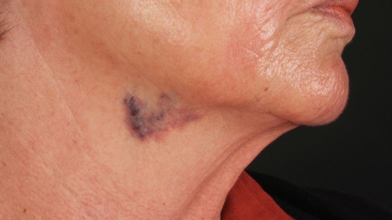
Cystic Swelling: Deep subcutaneous cystic swellings may occur, particularly in the neck, axilla, or groin.
-
Skin Lesions: Vesicles or other skin abnormalities may be noticed, especially in cases of lymphangioma circumscriptum.
Common Locations and Symptoms
Cavernous lymphangiomas can appear in various parts of the body, each with unique symptoms.
-
Head and Neck: These are the most common locations, particularly in the face, neck, and throat.
-
Trunk: Lesions can also occur on the chest and abdomen.
-
Extremities: They can appear on the arms and legs.
-
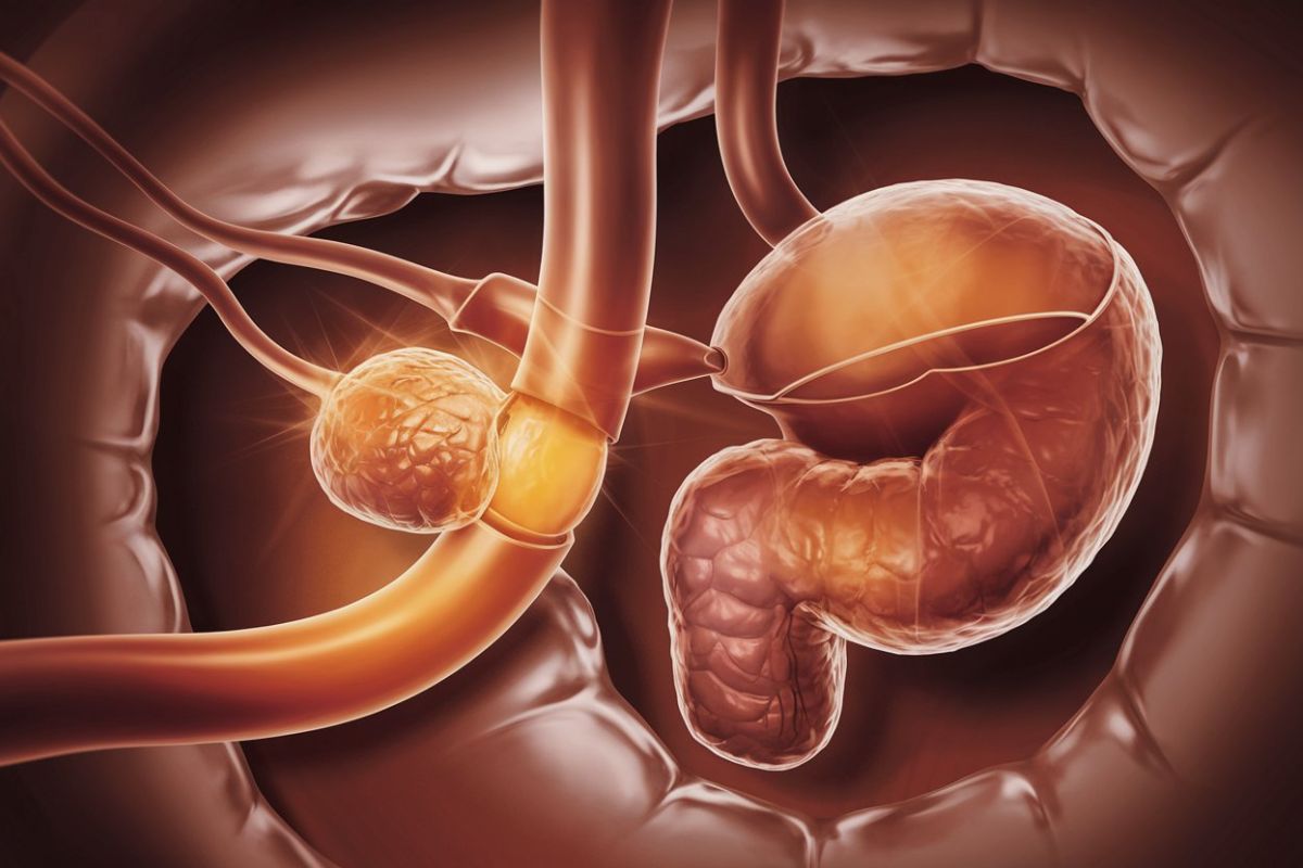
Internal Organs: Rarely, they can occur in internal organs such as the breast, liver, and small intestine.
-
Asymptomatic: Many lesions are asymptomatic and only discovered incidentally during medical examinations.
-
Minor Bleeding: Spontaneous episodes of minor bleeding may occur from ruptured vesicles.
-
Clear Fluid Drainage: Copious drainage of clear fluid from ruptured vesicles.
-
Respiratory Problems: Large cysts in the neck can cause dysphagia and respiratory problems.
-
Infection: Serious infections can occur if the lesions become infected.
Complications and Diagnosis
While generally benign, cavernous lymphangiomas can have serious complications. Accurate diagnosis is crucial for effective management.
-
Lymphangiosarcoma: A rare but aggressive malignancy that can arise from pre-existing lymphangiomas, particularly those exposed to radiation therapy.
-
Hemorrhage: Rarely, cavernous lymphangiomas can cause hemorrhage, especially in the scrotum.
-
Infection: Large cysts can become infected, leading to serious complications.
-
Ultrasonography: Useful for assessing the size and location of the lesion.
-
Mammography: Used for breast lesions.
-
Magnetic Resonance Imaging (MRI): Provides detailed images of the lesion and its relationship with surrounding tissues.
-
Histopathology: The final diagnosis is based on histological examination of tissue samples, which show dilated lymphatic channels lined by endothelial cells.
Treatment Options and Prognosis
Treatment depends on the size, location, and symptoms of the lesion. The prognosis is generally good, especially if the lesions are completely excised.
-
Surgical Excision: Complete surgical excision is often the preferred treatment for symptomatic lesions.
-
Observation: Small, asymptomatic lesions may be observed and treated only if they become symptomatic.
-
Radiation Therapy: Avoided due to the risk of lymphangiosarcoma.
-
Sclerotherapy: In some cases, sclerotherapy may be used to reduce the size of the lesion.
-
Prognosis: The prognosis for patients with cavernous lymphangiomas is generally good, especially if the lesions are completely excised. However, there is a risk of complications such as lymphangiosarcoma, especially if the lesion is exposed to radiation therapy.
Final Thoughts on Cavernous Lymphangioma
Cavernous lymphangioma, though rare, presents unique challenges in diagnosis and treatment. These benign malformations, characterized by abnormal lymphatic vessels, can appear anywhere from the skin to internal organs. Symptoms vary widely based on location and size, ranging from asymptomatic nodules to significant respiratory issues. Diagnosis often involves imaging techniques like ultrasonography and MRI, with histopathology confirming the condition. Treatment options include surgical excision, observation, and sclerotherapy, each chosen based on the lesion's characteristics. While generally benign, complications like infection and rare malignancies can occur. Understanding cavernous lymphangioma's complexities requires a multidisciplinary approach, involving surgeons, radiologists, and genetic counselors. Ongoing research and patient education are crucial for better management and outcomes. By staying informed and vigilant, healthcare providers can offer optimal care to those affected by this condition.
Frequently Asked Questions
Was this page helpful?
Our commitment to delivering trustworthy and engaging content is at the heart of what we do. Each fact on our site is contributed by real users like you, bringing a wealth of diverse insights and information. To ensure the highest standards of accuracy and reliability, our dedicated editors meticulously review each submission. This process guarantees that the facts we share are not only fascinating but also credible. Trust in our commitment to quality and authenticity as you explore and learn with us.