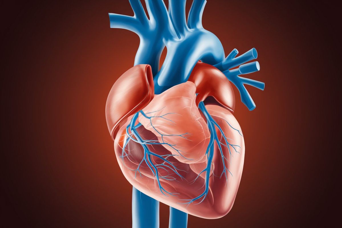
What is a cardiac diverticulum? Imagine a tiny pouch sticking out from the heart, containing all three layers of the heart wall: endocardium, myocardium, and pericardium. This rare condition, known as a cardiac diverticulum, can appear in different heart chambers like the left and right ventricles or the right atrium. While often harmless and discovered by accident during heart scans, these outpouchings can sometimes cause serious issues like arrhythmias, embolism, heart failure, or even rupture. Understanding the types, symptoms, and treatment options for cardiac diverticula is crucial for managing this unusual heart anomaly effectively.
Key Takeaways:
- Cardiac diverticula are rare outpouchings of the heart that can lead to serious complications like arrhythmias and heart failure. Early detection and appropriate treatment are crucial for improving outcomes.
- Imaging techniques such as echocardiography and MRI are essential for diagnosing and managing cardiac diverticula. Future research aims to improve diagnostic and treatment options for these rare heart anomalies.
What is a Cardiac Diverticulum?
Cardiac diverticula are rare outpouchings of the heart. They can be congenital or acquired and contain all three layers of the cardiac wall: endocardium, myocardium, and pericardium. Let's dive into some fascinating facts about these unique structures.
-
Definition: Cardiac diverticula are defined as congenital or acquired outpouchings of the heart that contain all three layers of the cardiac wall: endocardium, myocardium, and pericardium.
-
Types of Diverticula: There are several types of cardiac diverticula, including left ventricular (LV) diverticula, right ventricular (RV) diverticula, and right atrial (RA) diverticula. Each type has distinct characteristics and clinical implications.
-
Prevalence: Cardiac diverticula are rare, with a prevalence that varies depending on the type. For example, LV diverticula are estimated to occur in about 0.4% of cardiac autopsies.
Why Are They Clinically Significant?
Though often asymptomatic, cardiac diverticula can lead to serious complications. Understanding their clinical significance is crucial for proper management.
-
Clinical Significance: While most cardiac diverticula are asymptomatic, they can be associated with significant complications such as arrhythmias, embolism, heart failure, and rupture. These complications necessitate careful monitoring and appropriate treatment.
-
Symptoms: Symptoms of cardiac diverticula can vary widely and often depend on the size and location of the diverticulum. Common symptoms include chest pain, shortness of breath, and palpitations. In some cases, the diverticulum may be discovered incidentally during imaging studies for other conditions.
-
Diagnosis: Diagnosis of cardiac diverticula typically involves imaging techniques such as echocardiography, cardiac magnetic resonance imaging (MRI), and computed tomography (CT) angiography. These methods help in identifying the location and size of the diverticulum and assessing its impact on cardiac function.
Types of Cardiac Diverticula
Different types of cardiac diverticula have unique characteristics. Let's explore the specifics of each type.
-
Left Ventricular Diverticula: Left ventricular diverticula are the most common type of cardiac diverticula. They are often associated with other congenital anomalies and can be diagnosed during childhood. Most cases are asymptomatic but can lead to complications like arrhythmias and heart failure.
-
Right Ventricular Diverticula: Right ventricular diverticula are less common than LV diverticula and are often discovered incidentally during imaging studies for other conditions. They can be associated with arrhythmias and other cardiac anomalies.
-
Right Atrial Diverticula: Right atrial diverticula are rare and can be diagnosed with atrial arrhythmias early in life. They may remain asymptomatic until late in life and can be found incidentally during chest radiographs.
Potential Complications
Cardiac diverticula can lead to various complications. Understanding these risks is essential for effective management.
-
Complications: Complications of cardiac diverticula include arrhythmias, embolism, heart failure, and rupture. The risk of rupture is particularly high for large diverticula and can lead to life-threatening situations.
-
Arrhythmias: Arrhythmias are a common complication of cardiac diverticula. The diverticulum can act as a source of abnormal electrical activity, leading to various types of arrhythmias, including ventricular tachycardia.
-
Embolism: Embolism is another potential complication of cardiac diverticula. The diverticulum can serve as a source for thrombus formation, which can embolize and cause systemic or pulmonary embolism.
-
Heart Failure: Heart failure can occur in cases where the diverticulum significantly impairs cardiac function. The enlarged structure can compress adjacent cardiac chambers, leading to reduced cardiac output.
-
Rupture: Rupture of the diverticulum is a severe complication that can lead to life-threatening bleeding. It is more common in large diverticula and requires immediate surgical intervention.
Treatment Options
Treatment for cardiac diverticula varies based on size, location, and clinical significance. Here are some common approaches.
-
Treatment Options: Treatment options for cardiac diverticula depend on the size, location, and clinical significance of the diverticulum. Options include medical management, percutaneous closure, and surgical resection.
-
Medical Management: Medical management is often the first line of treatment for asymptomatic or small diverticula. This may involve monitoring with regular echocardiograms and other imaging studies to assess for any changes in the diverticulum.
-
Percutaneous Closure: Percutaneous closure is a minimally invasive procedure used to treat smaller diverticula. This involves using a catheter to deploy a device that closes the opening of the diverticulum.
-
Surgical Resection: Surgical resection is typically reserved for larger diverticula that pose a significant risk of rupture or other complications. The procedure involves removing the diverticulum and closing the opening to prevent further outpouching.
-
Surgical Approaches: Surgical approaches for resecting cardiac diverticula can vary depending on the location and size of the diverticulum. Common approaches include median sternotomy, thoracotomy, and minimally invasive techniques.
-
Postoperative Care: Postoperative care for patients undergoing surgical resection of cardiac diverticula is crucial to ensure proper healing and prevent complications. This includes monitoring for arrhythmias, managing pain, and ensuring adequate cardiac function.
Prognosis and Discovery
The prognosis for patients with cardiac diverticula can vary. Early detection and appropriate treatment are key to improving outcomes.
-
Prognosis: The prognosis for patients with cardiac diverticula varies widely depending on the size, location, and clinical significance of the diverticulum. Generally, early detection and appropriate treatment can significantly improve outcomes and prevent life-threatening complications.
-
Incidental Discovery: Many cardiac diverticula are discovered incidentally during imaging studies for other conditions. This highlights the importance of thorough imaging protocols to identify potential cardiac anomalies.
Genetic and Familial Factors
Genetic factors can play a role in the development of cardiac diverticula. Understanding these factors can help in early detection and management.
-
Family History: There is limited evidence suggesting a familial component in the development of cardiac diverticula. However, patients with a family history of cardiac anomalies may be at higher risk for developing similar conditions.
-
Genetic Factors: Genetic factors play a role in the development of some cardiac anomalies, including diverticula. Certain genetic syndromes can increase the risk of congenital heart defects, including those involving diverticula.
Special Cases: Prenatal and Pediatric
Cardiac diverticula can be diagnosed prenatally or in pediatric patients. Early detection is crucial for effective management.
-
Prenatal Diagnosis: Prenatal diagnosis of cardiac diverticula is rare but possible with advanced imaging techniques such as fetal echocardiography. Early detection can help in planning appropriate management strategies postnatally.
-
Pediatric Cases: Pediatric cases of cardiac diverticula are more common than adult cases due to the higher prevalence of congenital anomalies in children. Early detection and treatment are crucial to prevent long-term complications.
Adult Cases and Imaging Techniques
Adult cases of cardiac diverticula, though less common, can still pose significant challenges. Imaging techniques are essential for diagnosis and management.
-
Adult Cases: Adult cases of cardiac diverticula are less common but can still pose significant clinical challenges. These cases often present with symptoms such as chest pain and shortness of breath, necessitating prompt evaluation and treatment.
-
Imaging Techniques: Imaging techniques such as echocardiography, cardiac MRI, and CT angiography are essential for diagnosing and managing cardiac diverticula. These methods provide detailed information about the size, location, and impact of the diverticulum on cardiac function.
Therapeutic Strategies and Future Research
A multidisciplinary approach is often required for managing cardiac diverticula. Future research will help improve diagnostic and treatment options.
-
Therapeutic Strategies: Therapeutic strategies for cardiac diverticula involve a multidisciplinary approach, including cardiology, cardiothoracic surgery, and interventional cardiology. The choice of treatment depends on the specific characteristics of the diverticulum and the clinical condition of the patient.
-
Future Research: Future research in the field of cardiac diverticula should focus on improving diagnostic techniques, developing more effective treatment options, and understanding the underlying genetic and molecular mechanisms that contribute to their development. This will help in providing better management strategies for patients with these rare cardiac anomalies.
Final Thoughts on Cardiac Diverticulum
Cardiac diverticula, though rare, can have significant implications for heart health. These outpouchings, containing all three layers of the cardiac wall, can lead to complications like arrhythmias, embolism, heart failure, and even rupture. Diagnosis often involves imaging techniques such as echocardiography, MRI, and CT angiography. Treatment varies from medical management to surgical resection, depending on the diverticulum's size and location. Early detection and appropriate intervention are crucial for improving outcomes. Understanding the types, symptoms, and potential complications helps in managing this condition effectively. Future research aims to enhance diagnostic methods and treatment options, offering better care for those affected. Stay informed and consult healthcare professionals if you suspect any heart-related issues.
Frequently Asked Questions
Was this page helpful?
Our commitment to delivering trustworthy and engaging content is at the heart of what we do. Each fact on our site is contributed by real users like you, bringing a wealth of diverse insights and information. To ensure the highest standards of accuracy and reliability, our dedicated editors meticulously review each submission. This process guarantees that the facts we share are not only fascinating but also credible. Trust in our commitment to quality and authenticity as you explore and learn with us.


