


What exactly is a bronchogenic cyst? These rare congenital malformations arise from the abnormal budding of the tracheobronchial tree during early embryonic development. Typically found in the mediastinum or lung parenchyma, bronchogenic cysts often remain unnoticed until they cause symptoms like cough, fever, or chest pain. While many are asymptomatic, complications can occur if the cyst becomes infected or compresses nearby structures. Complete surgical removal is the recommended treatment to prevent potential issues. Understanding these cysts' origins, symptoms, and treatment options is crucial for effective management and ensuring a good prognosis. Dive into these 30 essential facts to learn more about bronchogenic cysts.
What Are Bronchogenic Cysts?
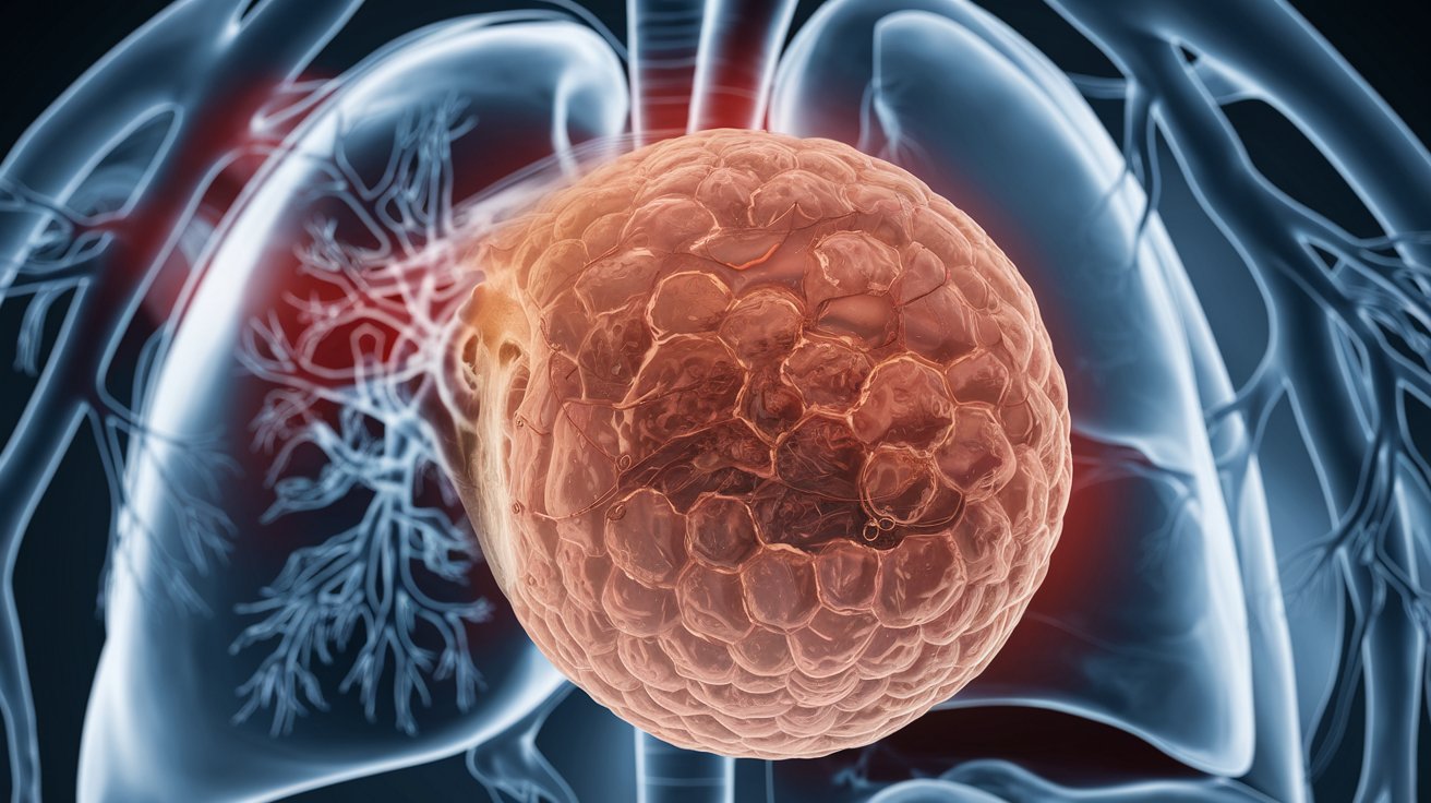
Bronchogenic cysts are rare congenital malformations of the respiratory tract. They arise from abnormal budding of the tracheobronchial tree during embryogenesis. Let's dive into some fascinating facts about these unique cysts.
-
Definition: Bronchogenic cysts are congenital malformations of the bronchial tree, a type of bronchopulmonary foregut malformation.
-
Etiology: They form due to abnormal budding of the embryonic ventral lung bud or the blind-ending pouch, which occurs between the 4th and 6th weeks of gestation.
-
Incidence: These cysts are rare, with a prevalence of 1 per 42,000 and 1 per 68,000 admissions in two hospital series.
Where Do Bronchogenic Cysts Occur?
These cysts can appear in various locations within the body. Their placement can significantly affect symptoms and treatment options.
-
Location: The most common location is the middle mediastinum (65-90%), but they can also be found in the lung parenchyma, particularly in the lower lobes.
-
Distribution: The distribution of bronchogenic cysts can vary widely, including locations such as the carinal area, paratracheal area, oropharyngeal wall, retrocardiac area, and intrapulmonary locations.
-
Intrapulmonary Cysts: Intrapulmonary cysts represent approximately 20% to 30% of all bronchogenic cysts and most commonly involve the lower lobes, but can also be found in the upper lobes.
-
Atypical Locations: Bronchogenic cysts can occur in atypical locations such as the neck, pericardium, pleura, diaphragm, and retroperitoneum, although these are rare.
Symptoms and Clinical Presentation
Bronchogenic cysts can be sneaky. Many people don't even know they have one until it causes problems.
-
Symptoms: Many bronchogenic cysts are asymptomatic and are discovered incidentally during imaging tests. However, symptoms can include cough, fever, pain, dyspnea, and hemoptysis when the cyst becomes infected or compresses adjacent structures.
-
Clinical Presentation: The clinical presentation can vary widely, from respiratory distress at birth to late appearance of symptoms. In pediatric patients, these cysts can cause life-threatening compressive symptoms, while in adults, they are often incidental radiologic findings.
-
Symptomatic Patients: Approximately 81% of patients with bronchogenic cysts are symptomatic, with cough being the most common symptom (45%).
Histopathology and Composition
Understanding the microscopic structure of bronchogenic cysts helps in diagnosing and treating them effectively.
-
Histopathology: The cysts are lined by secretory respiratory epithelium (cuboid or columnar ciliated epithelium) and contain fluid with variable amounts of proteinaceous material, blood products, and calcium oxalate.
-
Wall Composition: The cyst wall contains tissues similar to those of the normal bronchial tree, including cartilage, elastic tissues, mucous glands, and smooth muscle.
Size and Radiographic Features
The size and appearance of bronchogenic cysts on imaging tests can vary, making diagnosis a bit tricky.
-
Size: The size of bronchogenic cysts can vary from 2 to 12 cm in diameter, with most being unilocular and containing clear fluid.
-
Radiographic Features: On imaging, bronchogenic cysts typically appear as fluid-filled structures, but they can occasionally become air-filled following infection or intervention, resulting in an air-fluid level.
-
Plain Radiograph: The cysts may appear as spherical, smooth, white or pinkish masses with clear fluid, sometimes mimicking solid lesions due to the presence of calcium oxalate.
Complications and Treatment
Bronchogenic cysts can lead to various complications, especially if left untreated. Surgical intervention is often necessary.
-
Complications: Complications such as tracheobronchial compression, pulmonary infections, and rupture into the trachea, pericardial cavity, or pleural cavity can occur, especially in symptomatic patients.
-
Complications in Symptomatic Patients: Complications such as severe hemoptysis, pneumothorax, pleuritis, esophageal compression, infected cyst, and postobstructive pneumonia are more common in symptomatic patients.
-
Treatment: The treatment of all bronchogenic cysts is complete surgical excision, with definitive diagnosis established primarily by histopathological examination of the surgical specimen.
-
Surgical Approach: Surgical resection is recommended for all suspected bronchogenic cysts in operable candidates due to the risk of complications and the difficulty in making a confident preoperative diagnosis.
-
Postoperative Complications: Postoperative complications such as persistent air leak can occur but are relatively rare, with no late sequelae or recurrence reported in most cases.
-
Prognosis: The prognosis is excellent with complete resection, as there are no recurrences reported in case of complete surgical excision.
Bronchogenic Cysts in Adults and Children
Both adults and children can develop bronchogenic cysts, but their presentation and treatment might differ.
-
Incidence in Adults: Although bronchogenic cysts are more common in children, they can also be observed in adults, where they are often incidental radiologic findings.
-
Asymptomatic Cysts: Some bronchogenic cysts remain asymptomatic forever, but even these can eventually threaten life by producing compression, infection, hemorrhage, and rupture.
-
Symptoms Due to Infection: When bronchogenic cysts become infected, symptoms can include purulent sputum, fever, cough, and dyspnea, which force patients to seek medical attention.
Malignant Associations and Histological Findings
While rare, there are instances where bronchogenic cysts can be linked to more severe conditions.
-
Malignant Associations: There are rare instances where bronchogenic cysts are associated with malignancy, but this is not common and typically not a diagnostic concern.
-
Histological Findings: Histological examination of the surgical specimen reveals ciliated pseudostratified columnar epithelium of respiratory type, possible areas of squamous metaplasia, and the presence of airway components like cartilage plates and bronchial glands.
Study Findings and Radiological Features
Studies and imaging tests provide valuable insights into the nature of bronchogenic cysts.
-
Study Findings: A study of 12 patients with bronchogenic cysts found that six male and six female patients had an average age of 49 years, with six cases located in the pulmonary parenchyma and six in the mediastinum. The average size of the lesions was 5.3 cm, and most patients presented with cough, dyspnea, purulent sputum, fever, and asthenia.
-
Radiological Findings: Radiological findings include the presence of a cystic cavity with thin walls and hydro-aerial levels, which are often detected incidentally during routine imaging tests.
Importance of Infection
Infections can complicate bronchogenic cysts, making timely diagnosis and treatment crucial.
-
Importance of Infection: Infection is a significant complication of bronchogenic cysts, leading to purulent sputum and other symptoms that necessitate medical intervention. The study of infectious cases helps identify the microorganisms involved and guides appropriate treatment.
-
Clinical Characteristics: The clinical characteristics of patients with bronchogenic cysts include gender, age, diagnosis at admission, location, size, clinical condition, imaging studies, and the results of bacteriological cultures of the lesion content.
Final Thoughts on Bronchogenic Cysts
Bronchogenic cysts are rare congenital malformations that can cause a variety of symptoms, especially when infected or compressing nearby structures. These cysts, formed from abnormal budding of the embryonic ventral lung bud, are often found in the middle mediastinum or lung parenchyma. While many remain asymptomatic, others can lead to serious complications like tracheobronchial compression, infections, and even rupture. Surgical excision is the recommended treatment to prevent these issues and ensure a good prognosis. Understanding the clinical characteristics, radiological findings, and potential complications is crucial for accurate diagnosis and effective management. Despite their rarity, bronchogenic cysts can significantly impact a patient's health, making awareness and timely intervention essential.
Was this page helpful?
Our commitment to delivering trustworthy and engaging content is at the heart of what we do. Each fact on our site is contributed by real users like you, bringing a wealth of diverse insights and information. To ensure the highest standards of accuracy and reliability, our dedicated editors meticulously review each submission. This process guarantees that the facts we share are not only fascinating but also credible. Trust in our commitment to quality and authenticity as you explore and learn with us.