


What is Kasabach-Merritt Syndrome? Kasabach-Merritt Syndrome (KMS) is a rare, life-threatening condition that primarily affects infants. It involves a unique combination of blood clotting issues, severe thrombocytopenia (low platelet count), and vascular tumors like kaposiform hemangioendothelioma (KHE) or tufted angioma (TA). These tumors trap and consume platelets, leading to significant bleeding risks and complications. First identified in 1940, KMS presents with visible skin lesions, bruising, and sometimes internal bleeding. Early diagnosis and treatment are crucial for managing symptoms and improving outcomes. Understanding KMS can help in recognizing its signs and seeking timely medical intervention.
What is Kasabach-Merritt Syndrome?
Kasabach-Merritt Syndrome (KMS) is a rare and serious condition involving blood clotting issues due to specific vascular tumors. Understanding this syndrome is crucial for early diagnosis and treatment.
-
Definition and Etiology: Kasabach-Merritt Syndrome involves thrombocytopenia, consumptive coagulopathy, and purpura linked to kaposiform hemangioendothelioma (KHE) or tufted angioma (TA). These benign vascular tumors typically appear in infancy.
-
Historical Background: First described in 1940 by Haig Haigouni Kasabach and Katharine Krom Merritt, the syndrome was identified in a male infant with a large, rapidly growing lesion on his thigh.
Clinical Presentation and Symptoms
Recognizing the signs and symptoms of KMS can lead to timely medical intervention, potentially saving lives.
-
Common Signs and Symptoms: Visible cutaneous giant hemangiomas or multiple smaller hemangiomas, often on the extremities, enlarged abdomen, hepatomegaly or jaundice, petechiae, bruising, and frank bleeding are typical.
-
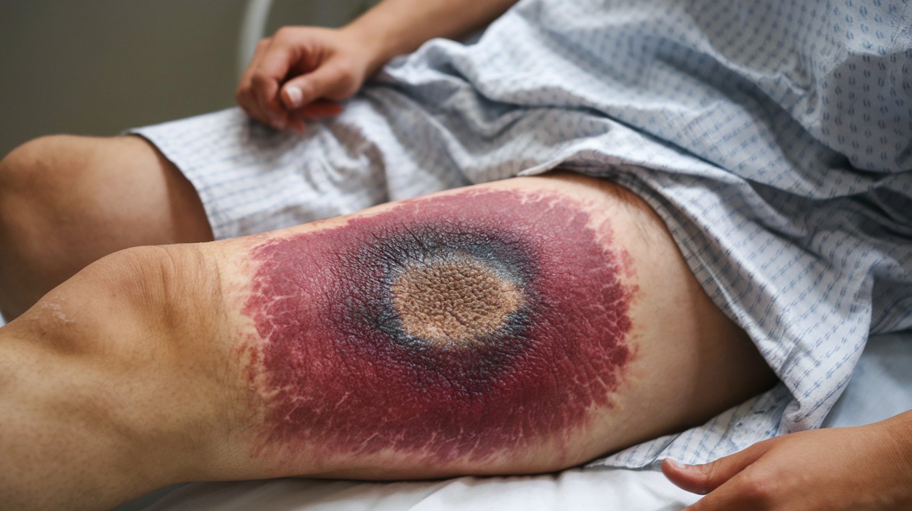
Physical Findings: Cutaneous hemangioma may appear as a large irregular bruise anywhere on the body. Kaposiform hemangioendothelioma or tufted angioma presents as blue or reddish-brown discoloration and skin induration. Petechiae, bruising, and bleeding from thrombocytopenia and coagulopathy are common.
Thrombocytopenia and Coagulopathy
Understanding the blood-related complications of KMS is essential for managing the condition effectively.
-
Thrombocytopenia: A hallmark of KMS, thrombocytopenia often presents with a platelet count less than 50,000/µL. Platelet trapping and aggregation within the vascular tumor lead to their consumption and subsequent low platelet count.
-
Consumptive Coagulopathy: Characterized by the consumption of clotting factors, leading to prolonged prothrombin time (PT) and activated partial thromboplastin time (aPTT), low fibrinogen levels, and elevated D-dimer levels. This can lead to disseminated intravascular coagulation (DIC), a significant cause of morbidity and mortality.
-
Microangiopathic Hemolytic Anemia: Another component of KMS, characterized by schistocytes (fragmented red blood cells) in the peripheral blood smear. This results from mechanical damage to red blood cells as they pass through the abnormal vascular channels within the tumor.
Vascular Tumors and Age of Onset
The types of tumors and the age at which KMS typically presents are critical for diagnosis.
-
Vascular Tumors: The two primary vascular tumors associated with KMS are kaposiform hemangioendothelioma (KHE) and tufted angioma (TA). These tumors are part of the same neoplastic spectrum but differ in clinical behavior.
-
Age of Onset: Most KMS cases occur in early infancy, with about 80% happening within the first year of life. Rare cases have been reported in the early neonatal period and even in adulthood, although these are extremely rare.
Location and Mortality Rate
The location of vascular lesions significantly impacts the prognosis and mortality rate of KMS.
-
Location of Vascular Lesions: Common sites include the face, head, neck, thoracic cavity, abdomen, retroperitoneum, and extremities. The location correlates with mortality rates, with skin tumors having a lower mortality rate compared to retroperitoneal tumors.
-
Mortality Rate: The overall mortality rate for KMS is suspected to be between 12 to 50%, with the most common causes of death being DIC-related hemorrhage, organ infiltration, high-output cardiac failure, multi-organ failure, or sepsis. Retroperitoneal tumors have a higher mortality rate due to their larger size and deeper location.
Diagnostic Evaluation and Imaging
Proper diagnosis and imaging are crucial for identifying and managing KMS.
-
Diagnostic Evaluation: Any infant with unexplained thrombocytopenia, with or without evidence of DIC, should be evaluated for visceral or hidden vascular lesions, especially of the liver or spleen. Laboratory studies include complete blood count (CBC) with differential, reticulocyte count, platelet count, peripheral smear examination, prothrombin time (PT), activated partial thromboplastin time (aPTT), fibrinogen, fibrin degradation product (FDP), and D-dimer levels.
-
Imaging Studies: Diagnostic imaging is crucial for identifying the extent of the vascular lesion and its impact on surrounding tissues. Imaging modalities such as radiography, computed tomography (CT), magnetic resonance imaging (MRI), and Doppler flow studies help assess the tumor's size and location.
Treatment Approaches
Managing KMS involves a combination of supportive therapy, medical treatment, and sometimes surgical intervention.
-
Treatment Approach: The primary goal is to control the coagulopathy and thrombocytopenia while managing the underlying tumor. Supportive therapy includes corticosteroids to reduce inflammation and sirolimus to inhibit cell proliferation. Once the coagulopathy stabilizes, surgical removal of the vascular tumor may be possible, although challenging due to the tumor's location and size.
-
Nonsurgical Treatment Regimens: Various nonsurgical treatments include systemic corticosteroids, irradiation, and various chemicals. The choice depends on the severity of the condition and the patient's overall health status.
-
Surgical Intervention: Surgery should be limited to symptomatic or complicated cases where other treatments have failed. Resection of the tumor is usually curative but can be difficult due to the tumor's location and the risk of bleeding complications.
Prognosis and Complications
Understanding the prognosis and potential complications of KMS is vital for patient care.
-
Prognosis: The prognosis varies widely depending on the location and size of the vascular lesion. Patients with skin tumors that undergo treatment have a lower mortality rate compared to those with retroperitoneal tumors.
-
Complications: High-output cardiac failure is a significant complication, leading to tachycardia, feeding difficulty, and shock. Multi-organ failure and sepsis are also potential complications due to the systemic nature of the disease.
Rare Cases and Genetic Considerations
Although primarily seen in infancy, KMS can occasionally present in adults, and genetic factors may play a role.
-
Rare Cases: While KMS is primarily associated with infancy, there have been reported cases in adulthood. For example, a 34-year-old female patient presented with multiple giant hepatic hemangiomas during pregnancy and multiple subcutaneous masses, managed with resection and embolization.
-
Genetic Considerations: There is no known ethnic predisposition to KMS, and both genders are equally affected. However, some cases may be associated with genetic syndromes, and chromosome tests may be necessary to rule out these conditions.
Understanding Kasabach-Merritt Syndrome
Kasabach-Merritt Syndrome (KMS) is a rare but serious condition linked to vascular tumors like kaposiform hemangioendothelioma and tufted angioma. It mainly affects infants, causing severe thrombocytopenia, consumptive coagulopathy, and sometimes life-threatening complications. Early diagnosis and treatment are crucial. Symptoms often include visible hemangiomas, bruising, and bleeding. Diagnostic tools like blood tests and imaging studies help identify the condition. Treatment usually involves managing the coagulopathy with medications like corticosteroids and sirolimus, and sometimes surgical removal of the tumor. Prognosis varies based on the tumor's location and size, with skin tumors generally having a better outcome than deeper, retroperitoneal ones. Ongoing research aims to improve treatment strategies and patient survival rates. Understanding KMS can lead to better management and outcomes for those affected by this challenging condition.
Was this page helpful?
Our commitment to delivering trustworthy and engaging content is at the heart of what we do. Each fact on our site is contributed by real users like you, bringing a wealth of diverse insights and information. To ensure the highest standards of accuracy and reliability, our dedicated editors meticulously review each submission. This process guarantees that the facts we share are not only fascinating but also credible. Trust in our commitment to quality and authenticity as you explore and learn with us.