
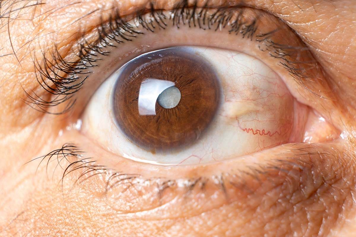

What is Iridocorneal Endothelial Syndrome (ICE)? Imagine waking up one day and noticing your vision isn't as clear as it used to be. You might be dealing with Iridocorneal Endothelial Syndrome, a rare eye disorder that affects the cornea, iris, and the fluid drainage system in your eye. ICE can cause symptoms like corneal swelling, secondary glaucoma, and changes in the iris. This condition usually affects women in their 30s to 50s and is often found in just one eye. Diagnosing ICE involves a thorough eye exam and specialized imaging techniques. Treatment can be tricky, often requiring surgery to manage glaucoma and corneal issues.
What is Iridocorneal Endothelial Syndrome?
Iridocorneal Endothelial Syndrome (ICE) is a rare eye disorder that affects the cornea, iris, and anterior chamber angle. It can cause significant vision problems and other complications. Let's dive into some key facts about this condition.
-
Definition and Classification
ICE syndrome includes three variants: Chandler syndrome, essential or progressive iris atrophy, and Cogan-Reese syndrome. Each has unique traits but shares the same underlying problem of abnormal corneal endothelial cell behavior. -
Clinical Features
Common symptoms include corneal edema, secondary glaucoma, iris atrophy, and unusual pupil shapes like distortion or multiple pupils (polycoria). These symptoms can severely impact vision. -
Pathophysiology
The condition involves normal corneal endothelial cells being replaced by cells that act more like epithelial cells. These new cells migrate and obstruct the iridocorneal angle, leading to the characteristic changes seen in ICE syndrome.
Corneal Edema and Secondary Glaucoma
Corneal edema and secondary glaucoma are significant issues in ICE syndrome. They contribute heavily to vision loss and require careful management.
-
Corneal Edema
Corneal edema results from elevated intraocular pressure (IOP) and poor pump function of the altered endothelial cells. This swelling can be severe and is a major cause of visual impairment. -
Secondary Glaucoma
Secondary glaucoma is common in ICE syndrome. The altered endothelium and high peripheral anterior synechiae (PAS) can close the angle, raising IOP and complicating diagnosis.
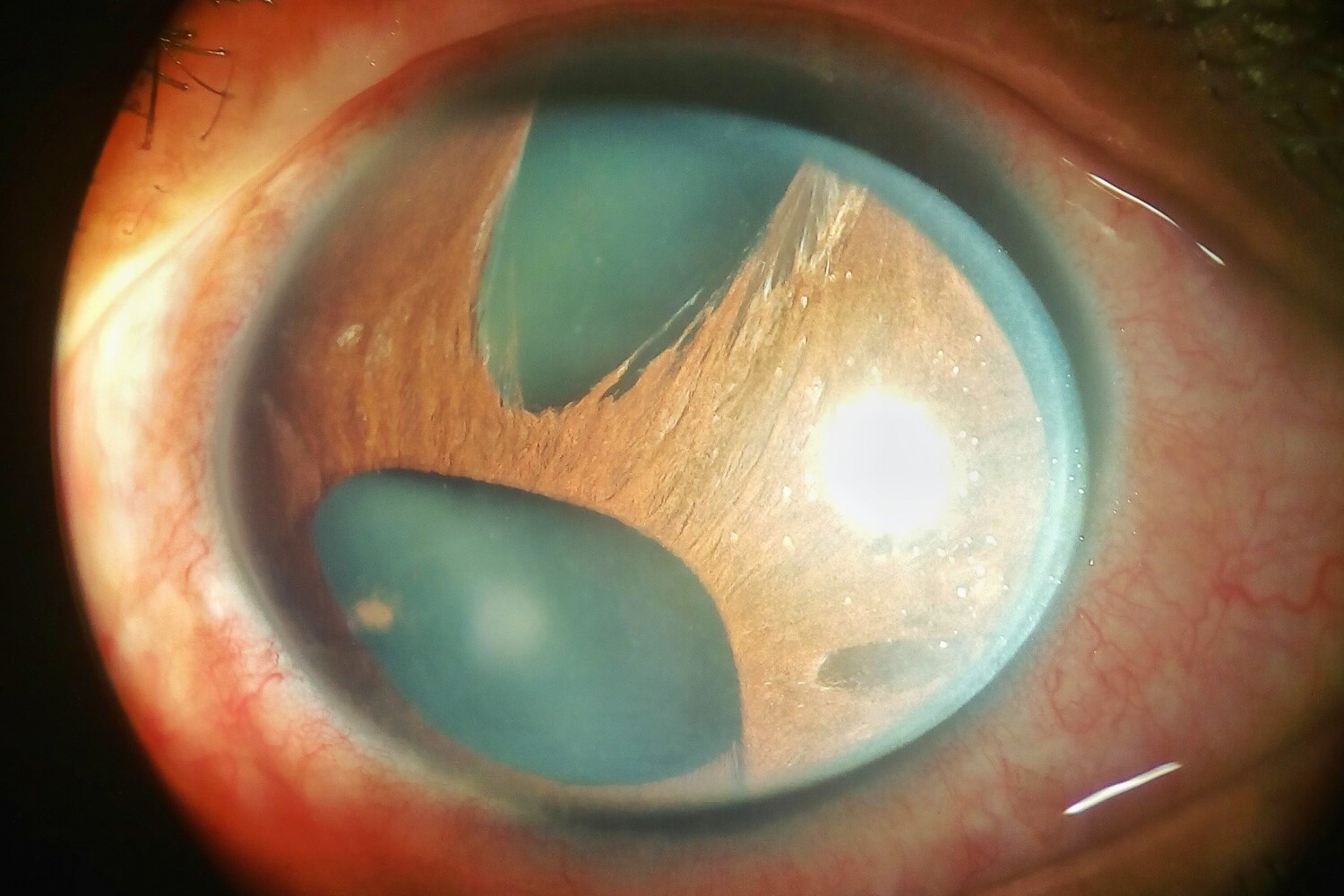
Iris Atrophy and Pupillary Anomalies
Changes in the iris and pupil are hallmark features of ICE syndrome. These alterations can be both diagnostic and symptomatic.
-
Iris Atrophy
Iris atrophy occurs due to the contraction of new tissue within the angle and on the iris. This leads to structural anomalies and further vision issues. -
Pupillary Anomalies
Distorted pupils or multiple pupils (polycoria) are common. These anomalies can significantly affect vision and help in diagnosing ICE syndrome.
Who is Affected?
Understanding who is most likely to develop ICE syndrome can help in early detection and management.
- Demographics
ICE syndrome usually affects women in their 30s to 50s. It is typically unilateral but can occasionally be bilateral.
Diagnosing ICE Syndrome
Accurate diagnosis is crucial for managing ICE syndrome effectively. Various techniques help in identifying this condition.
-
Diagnosis
A comprehensive eye exam, including tonometry and gonioscopy, is essential. Imaging techniques like in vivo confocal microscopy and ultrasound biomicroscopy confirm the diagnosis by revealing characteristic endothelial cell changes. -
Imaging Techniques
In vivo confocal microscopy is particularly useful. It visualizes the altered endothelial cells and their migration patterns, which are key indicators of ICE syndrome.
Differentiating ICE Syndrome from Other Conditions
Several other eye conditions can mimic ICE syndrome. Differentiating between them is vital for appropriate treatment.
-
Differential Diagnosis
Conditions like Cogan-Reese syndrome, Chandler syndrome, essential iris atrophy, posterior polymorphous corneal dystrophy (PPMD), and Axenfeld-Rieger syndrome need to be ruled out. Each has distinct features that help in differentiation. -
Posterior Polymorphous Corneal Dystrophy (PPMD)
PPMD is an inherited, bilateral condition that shares some features with ICE syndrome. However, it lacks the specific endothelial cell changes seen in ICE syndrome. -
Axenfeld-Rieger Syndrome (ARS)
ARS is congenital and bilateral, with systemic findings. Unlike ICE syndrome, ARS does not have the characteristic corneal endothelial changes.
Treatment Options
Managing ICE syndrome involves addressing both the glaucomatous and corneal aspects of the condition.
-
Treatment Options
Glaucoma filtering surgery with antifibrotic agents is often needed. Glaucoma drainage implants should be considered early to control IOP effectively. -
Endothelial Keratoplasty
This surgical procedure replaces the damaged endothelial layer with healthy donor tissue. It helps restore corneal clarity and improve vision.
Surgical Challenges and Prognosis
Surgery for ICE syndrome can be complex, and the prognosis varies depending on several factors.
-
Surgical Challenges
Surgical management is challenging due to the altered endothelium. Postoperative complications like persistent edema or recurrent glaucoma are common, requiring careful patient selection and technique. -
Prognosis
The prognosis depends on the severity and treatment effectiveness. Early diagnosis and appropriate management can improve outcomes, but some patients may still experience persistent visual impairment.
Potential Triggers and Bilateral Cases
Understanding potential triggers and monitoring for bilateral cases can aid in managing ICE syndrome.
-
Potential Triggers
The exact cause is unknown, but viral infections like herpes simplex virus (HSV) might trigger the condition. Studies have found HSV-DNA in many ICE syndrome cases. -
Bilateral Cases
While usually unilateral, ICE syndrome can be bilateral. Monitoring the other eye is crucial for early detection and management.
Comprehensive Management Strategies
A holistic approach is essential for managing ICE syndrome effectively.
- Management Strategies
A comprehensive approach addressing both glaucomatous and corneal aspects is necessary. This may involve medical therapy, surgical interventions, and supportive care. Regular follow-ups are essential to monitor progression and adjust treatment plans.
Final Thoughts on Iridocorneal Endothelial Syndrome
Iridocorneal Endothelial Syndrome (ICE) is a rare eye disorder that can seriously affect vision. With its three variants—Chandler syndrome, essential iris atrophy, and Cogan-Reese syndrome—ICE presents unique challenges. Key features include corneal edema, secondary glaucoma, and iris atrophy. Diagnosis often requires advanced imaging techniques like in vivo confocal microscopy. Treatment can be tricky, involving glaucoma surgeries and endothelial keratoplasty to manage symptoms and improve vision. While the exact cause remains unknown, potential triggers like viral infections have been suggested. Predominantly affecting women in their 30s to 50s, ICE is usually unilateral but can sometimes be bilateral. Early diagnosis and a comprehensive management strategy are crucial for better outcomes. Regular follow-ups help monitor disease progression and adjust treatments as needed. Understanding ICE's complexities can lead to more effective care and improved quality of life for those affected.
Was this page helpful?
Our commitment to delivering trustworthy and engaging content is at the heart of what we do. Each fact on our site is contributed by real users like you, bringing a wealth of diverse insights and information. To ensure the highest standards of accuracy and reliability, our dedicated editors meticulously review each submission. This process guarantees that the facts we share are not only fascinating but also credible. Trust in our commitment to quality and authenticity as you explore and learn with us.