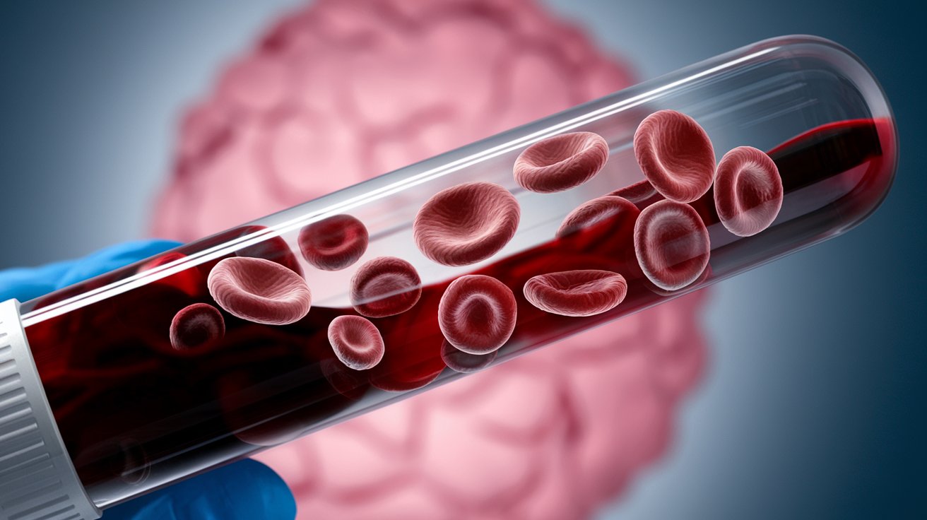
What is Hemoglobin Lepore Syndrome? Hemoglobin Lepore Syndrome is a rare genetic disorder affecting the blood. It results from a fusion of the delta and beta globin genes, leading to abnormal hemoglobin production. This condition can cause a range of health issues, from mild anemia to severe complications similar to beta-thalassemia. Found mostly in people of Balkan descent, it manifests differently depending on whether a person has one or two copies of the mutated gene. Those with one copy often show no symptoms, while those with two may suffer from severe anemia and related complications. Understanding this syndrome is crucial for proper diagnosis and treatment.
What is Hemoglobin Lepore Syndrome?
Hemoglobin Lepore syndrome is a rare genetic disorder affecting the hemoglobin in our blood. This condition can lead to various health issues, especially related to anemia. Let's dive into some key facts about this syndrome.
-
Definition and Cause: Hemoglobin Lepore syndrome is caused by a fusion of the delta and beta globin chains due to an unequal crossing over between the delta and beta globin genes.
-
Prevalence: This syndrome is relatively rare, with the highest prevalence found in populations of Balkan descent, particularly in Albania, Croatia, Serbia, Slovenia, and Romania.
Clinical Presentation and Diagnosis
Understanding how Hemoglobin Lepore syndrome presents itself and how it is diagnosed can help in managing the condition effectively.
-
Clinical Presentation: The homozygous state of Hb Lepore is rare and typically presents with severe anemia, splenomegaly, hepatomegaly, and skeletal abnormalities similar to those seen in beta-thalassemia major.
-
Heterozygous Form: Individuals who are heterozygous for Hb Lepore are generally asymptomatic and may present with mild microcytic hypochromic anemia, similar to the thalassemia trait.
-
Hemoglobin Levels: In heterozygotes, the hemoglobin level is typically within the normal range (11–13 g/dL), but there is significant hypochromia (deficiency of hemoglobin in the red blood cells) and microcytosis (small red blood cells).
-
Hemoglobin F (HbF): The presence of fetal hemoglobin (HbF) is increased in Hb Lepore syndrome, particularly in the homozygous state, where it can range from 3–5% of total hemoglobin.
-
Hemoglobin Electrophoresis: Hemoglobin electrophoresis is a crucial diagnostic tool for identifying Hb Lepore. It shows aberrant Hb Lepore fractions at a rate of 5–15% and a decreased percentage of HbA (normal adult hemoglobin).
-
Diagnosis: Diagnosis can be made through various tests including complete blood count (CBC), cation exchange high-performance liquid chromatography (CE-HPLC), hemoglobin electrophoresis, and DNA analysis.
Treatment and Management
Managing Hemoglobin Lepore syndrome involves understanding the treatment options and potential complications.
-
Treatment: Management of homozygous or compound heterozygous patients involves regular blood transfusions to alleviate severe anemia. Heterozygotes typically require no specific treatment.
-
Complications: A potential complication in children with severe anemia is the risk of silent stroke, which can cause brain damage without noticeable symptoms.
Epidemiology and Variants
Hemoglobin Lepore syndrome has different variants and is more common in certain populations. Let's explore these aspects.
-
Epidemiology: The Hb Lepore trait has a worldwide distribution but is more prevalent in specific ethnic groups, particularly Caucasians from Southern regions of Central and Eastern Europe.
-
Variants: There are three main variants of Hb Lepore: Washington (also known as Boston), Baltimore, and Hollandia. Each variant has distinct geographical and ethnic associations.
-
Washington Variant: The Washington variant is the most common and is prevalent in Italians from Southern Italy.
-
Baltimore Variant: The Baltimore variant is commonly found in people from the Balkan countries, including Albania, Croatia, Serbia, Slovenia, and Romania. It has also been reported in Turks and regions of Spain and Portugal.
-
Hollandia Variant: The Hollandia variant is identified in Papua New Guinea and Bangladesh.
Clinical Course and Associations
The clinical course of Hemoglobin Lepore syndrome can vary, and it can be associated with other conditions like beta-thalassemia.
-
Clinical Course: The clinical course of Hb Lepore syndrome can vary significantly. Patients with the homozygous state may present with severe anemia during the first two years of life, while heterozygotes are generally asymptomatic.
-
Thalassemia Association: Hb Lepore can be associated with beta-thalassemia, leading to a more severe clinical presentation. The combination of Hb Lepore with beta-thalassemia can result in transfusion-dependent anemia.
-
Iron Deficiency: Patients with Hb Lepore disease and associated beta-thalassemia are at risk for iron deficiency due to increased hemolysis. Therefore, assessing true iron status is crucial before considering iron supplements or transfusions.
Technological Advances in Diagnosis and Management
Modern technology plays a significant role in diagnosing and managing Hemoglobin Lepore syndrome.
-
Natural Language Processing (NLP): NLP can be used to automatically create enriched documents containing structured clinical information from patient reports. This integration with XML models provides efficient retrieval and highlighting of salient information.
-
Electronic Document Management: Electronic document management systems can improve procedure management in clinical laboratories by using markup languages like XML. A markup vocabulary such as CLP-ML (Clinical Laboratory Procedure Markup Language) supports various procedure types and enhances editing, reading, and searching capabilities.
Key Takeaways on Hemoglobin Lepore Syndrome
Hemoglobin Lepore syndrome, a rare genetic disorder, results from a fusion of delta and beta globin chains. Most common in Balkan populations, it can cause severe anemia, splenomegaly, and skeletal issues in its homozygous form. Heterozygous individuals usually show mild symptoms. Diagnosis involves blood tests and hemoglobin electrophoresis. Treatment for severe cases includes regular blood transfusions. There are three main variants: Washington, Baltimore, and Hollandia, each linked to specific regions. Increased fetal hemoglobin (HbF) levels and potential complications like silent strokes are notable. Understanding this condition's genetic and clinical aspects is crucial for effective management. Advanced diagnostic tools and electronic document systems can enhance patient care.
Was this page helpful?
Our commitment to delivering trustworthy and engaging content is at the heart of what we do. Each fact on our site is contributed by real users like you, bringing a wealth of diverse insights and information. To ensure the highest standards of accuracy and reliability, our dedicated editors meticulously review each submission. This process guarantees that the facts we share are not only fascinating but also credible. Trust in our commitment to quality and authenticity as you explore and learn with us.


