


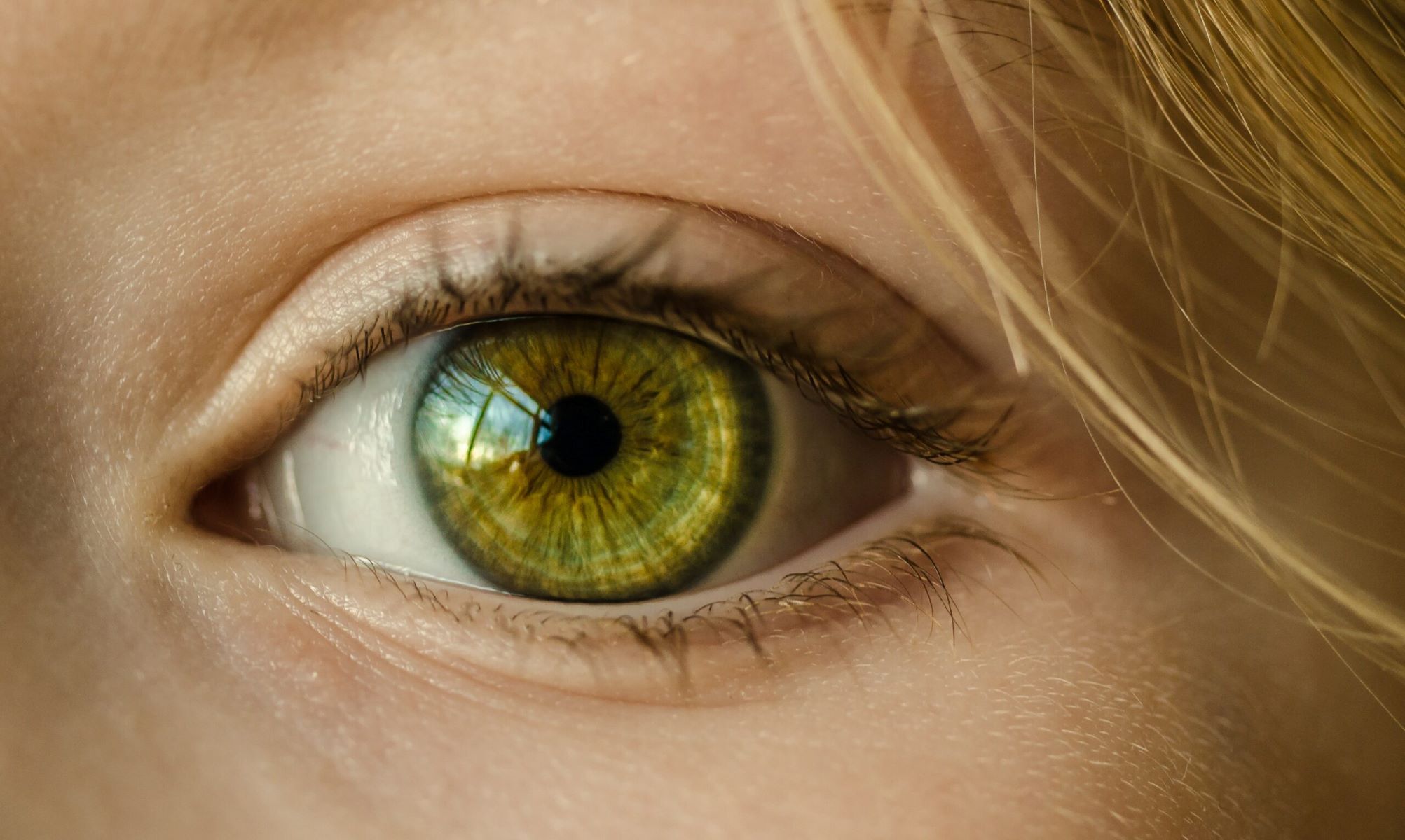
What is Foster–Kennedy Syndrome? Foster–Kennedy Syndrome (FKS) is a rare condition where one eye suffers optic atrophy while the other eye experiences papilledema. This happens due to an intracranial mass, often a meningioma, pressing on the optic nerve. The increased pressure in the brain causes swelling in the optic disc of the opposite eye. Patients might notice vision loss in one eye and headaches. Diagnosing FKS involves eye exams and imaging studies like CT scans or MRIs. Treatment usually requires a multidisciplinary approach, including surgery or radiation to address the underlying mass. Understanding FKS is crucial for timely and effective management.
What is Foster–Kennedy Syndrome?
Foster–Kennedy Syndrome (FKS) is a rare and complex condition affecting vision. It involves the optic nerves and is often linked to intracranial masses. Let's dive into some key facts about this intriguing syndrome.
-
Definition and Etiology
FKS occurs when an intracranial mass compresses the optic nerve on one side, causing optic atrophy. The increased intracranial pressure from the mass leads to papilledema in the opposite eye. -
Clinical Presentation
Patients typically present with unilateral visual loss due to optic atrophy in one eye and papilledema in the other. Central scotoma, or loss of vision in the middle of the visual field, may also occur.
Types of Foster–Kennedy Syndrome
FKS can be classified into different types based on the extent of optic nerve involvement and papilledema. Understanding these types helps in diagnosing and managing the condition.
-
Type 1
This type features optic atrophy in one eye and papilledema in the contralateral eye. -
Type 2
In Type 2, there is bilateral papilledema with unilateral optic atrophy. -
Type 3
This type involves bilateral papilledema that eventually leads to bilateral optic atrophy.
Common Causes and Risk Factors
Knowing the causes and risk factors can help in early detection and prevention of FKS.
-
Common Causes
The most frequent cause of FKS is an intracranial mass, especially meningiomas. These tumors often arise from the frontal lobe, olfactory groove, or sphenoid wing. -
Previous Radiation Exposure
Individuals who have received head radiation are at higher risk for developing meningiomas. -
Neurofibromatosis Type 2
This genetic disorder increases the likelihood of developing meningiomas. -
Female Gender
Women are more prone to developing meningiomas than men. -
Body Mass Index (BMI)
Higher BMI, waist circumference, and weight are associated with an increased risk of meningiomas.
Pathophysiology and Differential Diagnosis
Understanding the underlying mechanisms and differentiating FKS from similar conditions is crucial for accurate diagnosis.
-
Pathophysiology
FKS involves mechanical compression of the optic nerve by an intracranial mass, leading to optic atrophy. Increased intracranial pressure causes papilledema in the opposite eye. -
Differential Diagnosis
FKS can be mistaken for pseudo-Foster–Kennedy syndrome, which presents similarly but lacks an underlying compressive pathology. Other conditions include bilateral sequential anterior ischemic optic neuropathy (AION), idiopathic intracranial hypertension, and neurosyphilis.
Pseudo-Foster–Kennedy Syndrome
Pseudo-Foster–Kennedy Syndrome (PFKS) mimics FKS but has different underlying causes. Understanding PFKS helps in avoiding misdiagnosis.
-
Pseudo-Foster–Kennedy Syndrome
PFKS features unilateral optic atrophy with contralateral optic disc edema but lacks an underlying compressive pathology. The most common cause is bilateral sequential AION. -
Risk Factors for PFKS
Risk factors include hypertension, diabetes mellitus, high cholesterol, smoking history, and sleep apnea syndrome. -
Primary Prevention of PFKS
Controlling vasculopathic risk factors like blood pressure, blood sugars, and cholesterol levels can help prevent PFKS.
Diagnosis and Imaging Studies
Accurate diagnosis of FKS involves a thorough eye examination and imaging studies.
-
Diagnosis of FKS
Key findings include optic atrophy in the affected eye, papilledema in the contralateral eye, and central scotoma. Dilated fundoscopy is essential for adults with acute symptoms and vision loss. -
Imaging Studies
CT scans and MRIs help visualize the intracranial mass causing FKS. MRI provides detailed imaging of the brain and optic nerves.
Treatment and Follow-Up
Managing FKS requires a multidisciplinary approach and careful follow-up to ensure effective treatment.
-
Multidisciplinary Approach
Treatment involves neurologists, ophthalmologists, and radiologists. The primary goal is to manage the underlying intracranial mass and alleviate symptoms. -
Treatment Considerations
Surgical removal of the intracranial mass is often necessary. Radiation therapy and medications like corticosteroids may also be used. -
Follow-Up
Follow-up is crucial. Clinicians should monitor the affected optic nerve for four to eight weeks to ensure swelling is resolving and being replaced by pallor. Additional testing, including neuroimaging, should be ordered if there is any question about the etiology of PFKS.
Key Points on Foster–Kennedy Syndrome
Foster–Kennedy Syndrome (FKS) is a rare condition marked by unilateral optic atrophy and contralateral papilledema. It often stems from intracranial masses like meningiomas, primarily affecting the frontal lobe. Patients typically present with visual loss in one eye and papilledema in the other. Diagnosing FKS involves a thorough eye examination and imaging studies like CT scans and MRIs. Treatment usually requires a multidisciplinary approach, including surgery, radiation therapy, and medications.
Differentiating FKS from pseudo-Foster–Kennedy syndrome is crucial, as the latter lacks an underlying compressive pathology. Regular follow-ups are essential to monitor the condition and ensure effective management. Understanding the risk factors and pathophysiology of FKS can aid in early diagnosis and treatment, improving patient outcomes. Recognizing the symptoms and seeking prompt medical attention can make a significant difference in managing this complex syndrome.
Was this page helpful?
Our commitment to delivering trustworthy and engaging content is at the heart of what we do. Each fact on our site is contributed by real users like you, bringing a wealth of diverse insights and information. To ensure the highest standards of accuracy and reliability, our dedicated editors meticulously review each submission. This process guarantees that the facts we share are not only fascinating but also credible. Trust in our commitment to quality and authenticity as you explore and learn with us.