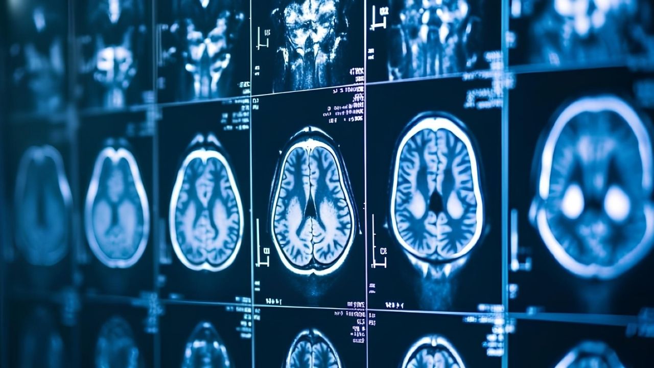
Neuroimaging has revolutionized our understanding of the brain. But what exactly is it? Neuroimaging refers to techniques that visualize the structure and function of the brain. These methods include MRI, CT scans, PET scans, and more. They help scientists and doctors see inside the brain without surgery. This technology has been crucial in diagnosing diseases like Alzheimer's, epilepsy, and brain tumors. It also aids in research on how the brain works, from memory to emotions. Ever wondered how we know which part of the brain lights up when you think? Neuroimaging is the answer. Dive into these 25 facts to learn more about this fascinating field!
Key Takeaways:
- Neuroimaging allows scientists to visualize the brain's structure and function using techniques like MRI and PET scans. It helps diagnose conditions and study brain development, but ethical concerns about privacy and accessibility need to be addressed.
- The history of neuroimaging is filled with milestones, from the first CT scan in 1971 to the development of fMRI in the 1990s. These advancements have transformed our understanding of the brain and its disorders, paving the way for modern diagnostic tools and research methods.
What is Neuroimaging?
Neuroimaging is a fascinating field that allows scientists to visualize the structure and function of the brain. This technology has revolutionized our understanding of the human mind. Here are some intriguing facts about neuroimaging.
- Neuroimaging techniques include MRI, fMRI, PET, and CT scans.
- MRI stands for Magnetic Resonance Imaging and uses magnetic fields to create detailed images of the brain.
- fMRI, or functional MRI, measures brain activity by detecting changes in blood flow.
- PET scans, or Positron Emission Tomography, use radioactive tracers to visualize brain function.
- CT scans, or Computed Tomography, use X-rays to create cross-sectional images of the brain.
Historical Milestones in Neuroimaging
The journey of neuroimaging has seen many significant milestones. These advancements have paved the way for modern techniques.
- The first CT scan was performed in 1971 by Sir Godfrey Hounsfield.
- MRI technology was developed in the early 1970s by Paul Lauterbur and Peter Mansfield.
- The first human PET scan was conducted in 1973.
- fMRI was introduced in the early 1990s, providing a non-invasive way to study brain activity.
- The Human Connectome Project, launched in 2009, aims to map the neural connections in the brain.
Applications of Neuroimaging
Neuroimaging has a wide range of applications in both clinical and research settings. These uses have transformed how we diagnose and treat brain disorders.
- Neuroimaging is used to diagnose conditions like Alzheimer's disease, epilepsy, and brain tumors.
- It helps in planning surgeries by providing detailed images of brain structures.
- Researchers use neuroimaging to study brain development and aging.
- It aids in understanding mental health disorders such as depression and schizophrenia.
- Neuroimaging is used in cognitive neuroscience to study how different brain regions are involved in various cognitive functions.
Technological Advancements in Neuroimaging
The field of neuroimaging is constantly evolving with new technologies and methods. These advancements have improved the accuracy and efficiency of brain imaging.
- High-resolution MRI scanners can now produce images with a resolution of less than 1 millimeter.
- Diffusion Tensor Imaging (DTI) is a type of MRI that maps the diffusion of water molecules in brain tissue, revealing the brain's white matter tracts.
- Magnetoencephalography (MEG) measures the magnetic fields produced by neural activity, providing millisecond-level temporal resolution.
- Advanced PET scanners can now detect smaller amounts of radioactive tracers, reducing the radiation dose to patients.
- Machine learning algorithms are being developed to analyze neuroimaging data and identify patterns associated with specific brain disorders.
Ethical Considerations in Neuroimaging
With great power comes great responsibility. The use of neuroimaging raises several ethical questions that need to be addressed.
- Privacy concerns arise from the ability to potentially read thoughts or intentions from brain scans.
- The use of neuroimaging in legal settings, such as lie detection, raises questions about its reliability and ethical implications.
- There are concerns about the potential misuse of neuroimaging data for purposes like neuromarketing or surveillance.
- Informed consent is crucial when conducting neuroimaging studies, especially with vulnerable populations like children or individuals with mental health disorders.
- The cost and accessibility of neuroimaging technology can create disparities in healthcare, with some populations having limited access to these advanced diagnostic tools.
The Power of Neuroimaging
Neuroimaging has revolutionized our understanding of the brain. From MRI and CT scans to PET and fMRI, these technologies offer a window into the mind's inner workings. They help diagnose conditions like Alzheimer's, stroke, and brain tumors. Researchers use them to study everything from cognitive functions to mental health disorders.
Neuroimaging also plays a crucial role in neurosurgery, guiding surgeons with pinpoint accuracy. It aids in drug development, allowing scientists to see how medications affect brain activity. The field continues to evolve, promising even more insights and applications in the future.
Understanding neuroimaging's impact helps us appreciate its role in medicine and research. It's a powerful tool that continues to shape our knowledge of the brain, offering hope for better treatments and deeper understanding of human cognition.
Frequently Asked Questions
Was this page helpful?
Our commitment to delivering trustworthy and engaging content is at the heart of what we do. Each fact on our site is contributed by real users like you, bringing a wealth of diverse insights and information. To ensure the highest standards of accuracy and reliability, our dedicated editors meticulously review each submission. This process guarantees that the facts we share are not only fascinating but also credible. Trust in our commitment to quality and authenticity as you explore and learn with us.
