


What is a compound microscope? A compound microscope is a powerful tool that uses multiple lenses to magnify tiny objects, making them visible to the human eye. Unlike simple microscopes, which use just one lens, compound microscopes combine two or more lenses to achieve higher magnification. This allows scientists, students, and hobbyists to explore the intricate details of cells, bacteria, and other microscopic entities. Compound microscopes are essential in fields like biology, medicine, and materials science. They have revolutionized our understanding of the microscopic world, enabling discoveries that were once impossible. Whether you're a budding scientist or just curious about the unseen world, learning about compound microscopes can open up a whole new perspective.
What is a Compound Microscope?
A compound microscope is a powerful tool used to magnify small objects that are invisible to the naked eye. It uses multiple lenses to achieve high levels of magnification, making it essential in fields like biology, medicine, and materials science.
- The compound microscope was invented in the late 16th century by Dutch spectacle makers Hans and Zacharias Janssen.
- It uses two sets of lenses: the objective lens and the eyepiece lens, to magnify objects.
- The term "compound" refers to the microscope's use of multiple lenses to achieve higher magnification.
- Compound microscopes can magnify objects up to 1,000 times their original size.
- The eyepiece lens typically magnifies objects 10 times, while objective lenses can vary from 4x to 100x magnification.
- The total magnification is calculated by multiplying the magnification of the eyepiece lens by that of the objective lens.
How Does a Compound Microscope Work?
Understanding how a compound microscope works can help you appreciate its complexity and usefulness. It involves a series of lenses and light sources to produce a clear, magnified image.
- Light from a source, such as a mirror or built-in lamp, passes through the specimen.
- The objective lens, located near the specimen, magnifies the image.
- This magnified image is then further enlarged by the eyepiece lens.
- The final image is viewed through the eyepiece, appearing much larger than the actual specimen.
- Fine and coarse focus knobs adjust the distance between the lenses and the specimen for a clear image.
- The stage holds the specimen slide in place and can be moved vertically or horizontally.
Types of Compound Microscopes
There are various types of compound microscopes, each designed for specific applications. Knowing these types can help you choose the right microscope for your needs.
- The brightfield microscope is the most common type, using light to illuminate the specimen.
- Darkfield microscopes enhance contrast in unstained specimens by using a special condenser.
- Phase-contrast microscopes allow for the study of transparent specimens without staining.
- Fluorescence microscopes use high-intensity light to excite fluorescent molecules in the specimen.
- Polarizing microscopes are used to study specimens that are birefringent, such as crystals and minerals.
- Confocal microscopes use laser light to scan specimens and produce high-resolution images.
Applications of Compound Microscopes
Compound microscopes are used in various fields, from education to advanced scientific research. Their versatility makes them indispensable in many areas.
- In biology, they are used to study cells, tissues, and microorganisms.
- Medical laboratories use them for diagnosing diseases by examining blood, tissues, and other samples.
- Forensic scientists use compound microscopes to analyze evidence such as hair, fibers, and residues.
- In materials science, they help in examining the microstructure of metals, polymers, and ceramics.
- Environmental scientists use them to study microorganisms in water and soil samples.
- Educational institutions use compound microscopes to teach students about the microscopic world.
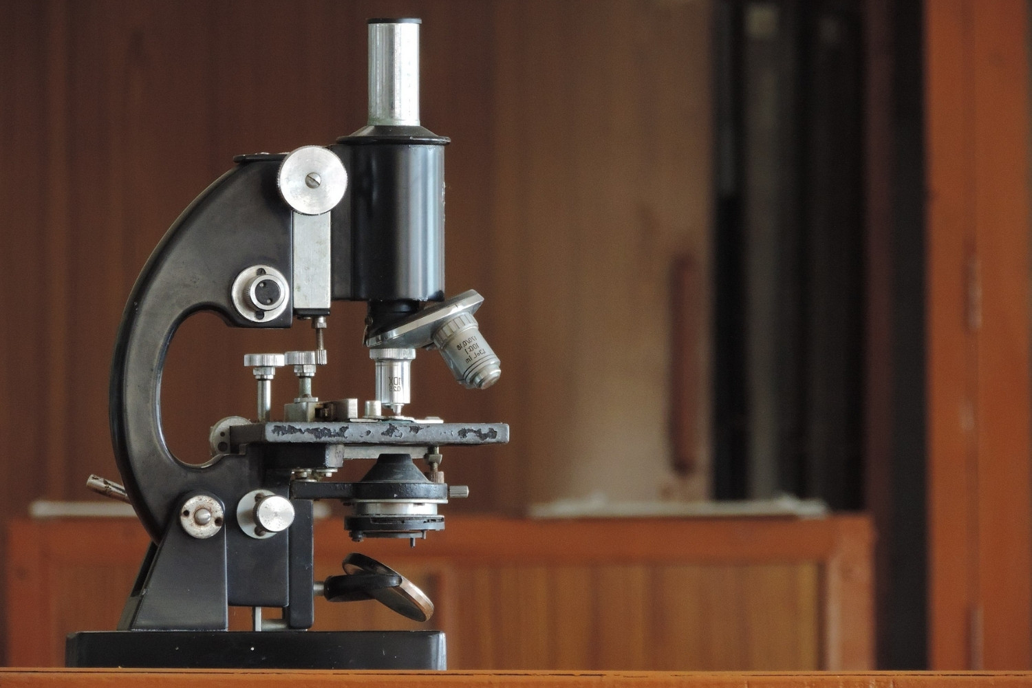
Key Components of a Compound Microscope
A compound microscope consists of several key components, each playing a crucial role in its function. Understanding these parts can help you use the microscope more effectively.
- The eyepiece, or ocular lens, is where you look through to see the magnified image.
- Objective lenses are located on a rotating nosepiece and provide different levels of magnification.
- The stage is a flat platform where the specimen slide is placed.
- Stage clips or a mechanical stage hold the slide in place.
- The light source, such as a mirror or built-in lamp, illuminates the specimen.
- The condenser focuses light onto the specimen for better illumination.
- Coarse and fine focus knobs adjust the focus of the image.
- The arm connects the base to the head and is used for carrying the microscope.
- The base provides stability and supports the entire microscope.
Interesting Facts About Compound Microscopes
Here are some fascinating facts that highlight the importance and versatility of compound microscopes.
- The first compound microscopes were very crude and had limited magnification.
- Antonie van Leeuwenhoek, known as the "Father of Microbiology," made significant improvements to the microscope.
- Modern compound microscopes can achieve magnifications of up to 2,000 times.
- Electron microscopes, which are a type of advanced compound microscope, can magnify objects up to 10 million times.
- Compound microscopes have played a crucial role in many scientific discoveries, including the identification of cells and bacteria.
The Final Lens
Compound microscopes have revolutionized how we see the microscopic world. From their invention to modern advancements, these tools have opened up new realms of discovery. They’ve played a crucial role in medicine, biology, and countless other fields. Understanding their parts and functions can deepen appreciation for their impact.
Whether you're a student, a scientist, or just curious, knowing these facts can enhance your grasp of the microscopic universe. The next time you peer through a microscope, remember the rich history and intricate technology behind it.
These instruments aren’t just tools; they’re gateways to understanding life at its most fundamental level. So, keep exploring, keep learning, and let your curiosity guide you. The microscopic world is vast and full of wonders waiting to be discovered. Happy exploring!
Was this page helpful?
Our commitment to delivering trustworthy and engaging content is at the heart of what we do. Each fact on our site is contributed by real users like you, bringing a wealth of diverse insights and information. To ensure the highest standards of accuracy and reliability, our dedicated editors meticulously review each submission. This process guarantees that the facts we share are not only fascinating but also credible. Trust in our commitment to quality and authenticity as you explore and learn with us.